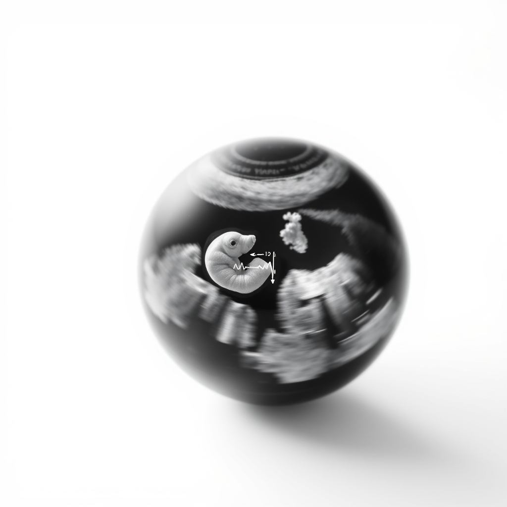
Ultrasounds are done 2-3 times in a normal pregnancy, how often ultrasounds normal pregnancy depends on the stage and health of the fetus and mother
By Shubhra Mishra — a mom of two who turned her own confusion during pregnancy into BumpBites, a global mission to make food choices clear, safe, and stress-free for every expecting mother. 💛
Check whether any food is safe during pregnancy with the BumpBites Food Safety Checker.
Quick take: Most low‑risk pregnancies have 2–3 routine ultrasounds—one early dating scan, a detailed anatomy scan around 20 weeks, and an optional growth scan later on. They are considered safe when performed by qualified providers, and extra scans are only recommended if specific medical concerns arise.
It’s 2 a.m., you’re lying in bed, and a sudden cramp makes you wonder: “Did I just miss the perfect window for an ultrasound?” You’re not alone. Many expecting parents worry about how often they should schedule scans, whether they’re safe, and what each appointment actually looks like. The good news is that the answer isn’t a one‑size‑fits‑all number—guidelines balance the benefits of seeing your baby’s growth with the principle of using the lowest effective exposure.
In this article we’ll walk through the typical ultrasound schedule, explain why each scan matters, clarify safety limits, and help you decide when an extra scan might be appropriate. We’ll also cover insurance basics, preparation tips, and alternatives if you’re looking for other ways to monitor your pregnancy. By the end you’ll have a clear, evidence‑based roadmap for “how often ultrasounds normal pregnancy” and feel confident discussing it with your provider.
How many ultrasounds are recommended during a normal pregnancy?
For a low‑risk, uncomplicated pregnancy, most professional societies—including the American College of Obstetricians and Gynecologists (ACOG) and the UK's National Institute for Health and Care Excellence (NICE)—recommend 2 to 3 routine ultrasounds. The first is a dating scan (usually 8–12 weeks), the second is a detailed anatomy scan (around 18–22 weeks), and a third—often optional—is a growth scan in the third trimester (around 28–32 weeks) if there are concerns about fetal size or maternal health.
These three scans provide key information:
Dating scan: Confirms gestational age, checks for a single versus multiple pregnancy, and rules out early complications.
Anatomy scan: Evaluates organ development, detects major structural anomalies, and confirms placental location.
Growth scan: Assesses fetal size, amniotic fluid volume, and placental health, especially if the mother has hypertension, diabetes, or a history of growth concerns.
In many countries, insurance plans (public or private) cover these three scans as part of standard prenatal care. Adding more scans without a clear medical indication is generally not covered and may not provide additional benefit.
For high‑risk pregnancies—such as those with maternal hypertension, gestational diabetes, or a known fetal condition—additional targeted scans are often ordered. The exact number then depends on the specific condition and the provider’s monitoring plan.
Recent ACOG updates emphasize that the timing of each scan can be individualized. For example, if a first‑trimester scan suggests a borderline fetal size, a follow‑up growth assessment may be scheduled earlier than the typical 28‑week window. This flexibility helps clinicians tailor care while still respecting the overall safety framework.
When should the first ultrasound be done in a healthy pregnancy?
The earliest routine scan is the dating ultrasound, typically performed between 8 and 12 weeks gestation. This window offers a clear view of the gestational sac, yolk sac, and often the embryo’s heartbeat. Doing the scan within this window provides the most accurate estimate of the due date, which guides subsequent prenatal visits and testing.
If you missed the 8–12‑week window, an ultrasound can still be useful later in the first trimester (up to 14 weeks). However, the crown‑rump length (CRL) measurement becomes less precise after 13 weeks, and the margin of error for dating widens.
Some parents opt for an even earlier “early pregnancy” scan at 6–7 weeks to confirm a viable intrauterine pregnancy after a positive home test. While technically possible, most guidelines advise reserving the earliest scan for 8 weeks to avoid unnecessary exposure and to ensure the embryo is large enough for reliable imaging.
During the dating scan, the sonographer will also check for a single gestation, the position of the placenta, and any early signs of ectopic pregnancy. If any concerns arise, your provider may schedule a follow‑up scan or additional testing.
Because early dating is crucial for planning later screening (such as cell‑free DNA testing), many clinicians encourage patients to book the scan as soon as they have a reliable last menstrual period (LMP) date. This proactive approach can reduce uncertainty and streamline the overall prenatal timeline.
Typical dating scan at 10 weeks shows the embryo’s heartbeat and helps lock in your due date.
How often should I get an ultrasound in the second trimester?
The second trimester is when the anatomy scan (also called the level‑II scan) is scheduled. Most guidelines place this appointment between 18 and 22 weeks, with 20 weeks being the most common target. This timing balances fetal size—large enough to see most organs—with the mother’s comfort, as the uterus is still relatively low.
During the anatomy scan, the sonographer assesses:
Brain and spinal cord development
Heart chambers and rhythm
Kidneys, bladder, and gastrointestinal tract
Lungs, limbs, and facial features
Placental position and umbilical cord insertion
If there are maternal risk factors (e.g., diabetes, hypertension, or a family history of congenital anomalies), the provider may schedule an additional targeted scan a few weeks later to monitor specific concerns.
Some parents ask whether a second‑trimester “growth scan” is needed. While growth assessment can be done in the third trimester, a mid‑second‑trimester scan is rarely required unless there are particular red‑flag concerns identified earlier.
Overall, for a low‑risk pregnancy, one comprehensive anatomy scan in the second trimester is sufficient. The scan usually lasts 20‑30 minutes, and you’ll leave with printed images or a digital copy to share with family.
In addition to the standard anatomy scan, many clinics now offer optional “soft‑tissue” assessments, such as fetal facial profile or limb measurements, which can provide extra reassurance without significantly extending exposure time. Discuss these options with your provider if you’re curious.
Are routine ultrasounds safe for the baby and mother?
Yes—when performed by trained professionals using the lowest effective settings, routine obstetric ultrasounds are considered safe for both the fetus and the mother. The U.S. Food and Drug Administration (FDA) classifies diagnostic ultrasound as a Class II medical device, meaning it is “generally recognized as safe when used appropriately.” The World Health Organization (WHO) and ACOG echo this, emphasizing that there is no evidence of harm at standard exposure levels.
Ultrasound works by sending high‑frequency sound waves into the body and measuring the echoes that return. Unlike ionizing radiation (e.g., X‑rays), sound waves do not carry enough energy to cause DNA damage. However, the principle of “ALARA”—as low as reasonably achievable—still applies. This means the sonographer will use the lowest power needed to obtain a clear image, limit the duration of exposure, and avoid unnecessary scans.
Most safety guidelines recommend keeping total exposure under 30 minutes per pregnancy, although typical routine scans total far less than 10 minutes of active imaging time. Studies involving thousands of pregnancies have not shown a link between standard obstetric ultrasound and adverse outcomes such as birth defects, neurodevelopmental problems, or growth restriction.
That said, certain cautions remain:
Do not use ultrasound for non‑medical “keepsake” videos or to determine the baby’s gender before the recommended window (usually 18 weeks).
Avoid prolonged “real‑time” scanning without a clear clinical purpose.
Pregnant women with specific medical implants (e.g., certain cardiac devices) should discuss any potential interaction with their provider, although most modern devices are unaffected.
If you have concerns about safety, ask your sonographer about the machine’s settings and the specific purpose of the scan. Transparency helps maintain trust and ensures you stay within recommended exposure limits.
Emerging research into the thermal effects of high‑intensity focused ultrasound (HIFU) is ongoing, but standard obstetric scanners operate well below those thresholds, reinforcing the consensus that routine imaging is safe.
What is the difference between routine and diagnostic ultrasounds in pregnancy?
Routine ultrasounds are scheduled as part of standard prenatal care for low‑risk pregnancies. Their primary goals are dating, anatomical assessment, and growth monitoring. They follow a predictable schedule, are covered by most insurance plans, and are performed using standard settings.
Diagnostic ultrasounds are ordered when a specific clinical question arises—such as checking for a suspected anomaly, evaluating fetal position after a breech presentation, or monitoring a known condition like twin‑twin transfusion syndrome. These scans may use higher‑resolution probes, longer observation periods, and sometimes employ Doppler techniques to assess blood flow.
Key distinctions:
Aspect
Routine Ultrasound
Diagnostic Ultrasound
Purpose
Standard dating, anatomy, growth
Targeted assessment of a specific concern
Frequency
2‑3 times per pregnancy
As needed; may be multiple in a short period
Insurance coverage
Usually covered
Often covered when medically indicated
Duration
10‑30 minutes
15‑45 minutes or longer
Technique
Standard B‑mode imaging
May include Doppler, 3‑D/4‑D, or specialized modes
Because diagnostic scans focus on a particular issue, they may be repeated more frequently if the condition evolves. For example, a mother with gestational diabetes might have weekly growth scans to track fetal size. In contrast, routine scans are spaced out to avoid unnecessary exposure while still providing essential information.
Both types are safe when performed correctly, but the diagnostic category often involves more detailed imaging, so the provider will again follow the ALARA principle to keep exposure as low as possible.
Can I have too many ultrasounds during pregnancy?
While there is no strict “maximum number,” exceeding the recommended amount of routine scans without a clear medical reason can be unnecessary. The cumulative exposure from many scans is still far below levels shown to cause harm, but unnecessary imaging adds cost, takes time, and may cause anxiety.
Research from the National Institute of Environmental Health Sciences (NIEHS) and other groups indicates that routine obstetric ultrasound exposure—well under 30 minutes total—does not increase the risk of miscarriage, low birth weight, or neurodevelopmental issues. However, a small number of studies have raised theoretical concerns about repeated high‑intensity exposure, especially in the first trimester, prompting the “as low as reasonably achievable” guideline.
Practical advice:
Stick to the standard 2‑3 scans unless your provider identifies a specific reason for additional imaging.
If you’re pregnant with twins or have a high‑risk condition, follow the schedule your provider recommends—these plans are designed to balance benefit and safety.
Avoid “keepsake” scans purely for entertainment; they often extend exposure without a medical need.
In short, for most low‑risk pregnancies, the routine schedule is sufficient. If you’re considering extra scans, discuss the reason with your obstetrician or midwife to ensure the benefit outweighs any theoretical risk.
Some clinics now offer “limited‑view” scans for non‑clinical purposes that use lower acoustic power. While technically safer, they still occupy clinic time and may reduce availability for patients who need medically indicated scans.
What factors affect the frequency of ultrasounds in pregnancy?
Several variables influence how many scans you’ll have:
Maternal health conditions: Hypertension, diabetes, obesity, or infections may prompt more frequent monitoring.
Fetal factors: Multiple gestations, abnormal growth patterns, or detected anomalies often require additional targeted scans.
Maternal age and history: Women over 35 or those with a previous pregnancy loss may be offered extra reassurance scans.
Provider practice style: Some obstetricians prefer a more hands‑on approach and schedule additional scans for reassurance, while others limit imaging to the three‑scan standard.
Insurance coverage: In the U.S., many private insurers cover three routine scans; any extra scans usually need a prior authorization or may be out‑of‑pocket.
Geographic guidelines: The NHS in the UK typically offers two scans—dating at 12 weeks and anatomy at 20 weeks—while ACOG in the U.S. recommends three, reflecting slight regional differences.
Understanding these factors helps you anticipate why your provider might suggest more or fewer scans. If you feel uncertain, ask for a clear explanation: “What specific aspect of my pregnancy are we monitoring with this extra scan?” This conversation can clarify the medical rationale and ensure you’re comfortable with the plan.
It’s also worth noting that lifestyle factors, such as smoking or nutrition, can influence the need for additional monitoring. Providers may adjust the ultrasound schedule based on how these factors affect fetal growth trajectories.
Seeing the baby's heartbeat on a routine scan can turn anxiety into excitement.
How to prepare for a prenatal ultrasound and what to expect during each appointment?
Preparation varies slightly by trimester:
First‑trimester dating scan: You’ll usually be asked to have a full bladder (about 1‑2 cups of water) 30 minutes before the appointment. A full bladder lifts the uterus, giving a clearer view of the gestational sac.
Second‑trimester anatomy scan: A moderately full bladder is still helpful, but many clinics no longer require it if you’re comfortable. Wear a two‑piece outfit if possible—this makes it easier for the sonographer to place the transducer.
Third‑trimester growth scan: A half‑full bladder is typically sufficient. Bring a list of any new symptoms (e.g., decreased fetal movement) to discuss.
During the scan, you’ll lie on an exam table while a gel is applied to your abdomen. The gel helps transmit sound waves and feels cool, not cold. The sonographer moves a small handheld probe over your belly, capturing real‑time images on a monitor. You may be asked to hold your breath briefly to improve image clarity.
Most scans last 15‑30 minutes. You’ll receive printed images or a digital file, and the sonographer will point out key structures (e.g., the head, heart, spine). If anything unusual is noted, the provider will discuss next steps—often simply a follow‑up scan.
Alternative monitoring methods include:
Doppler fetal monitoring: Uses sound waves to assess blood flow and heart rate, useful for high‑risk pregnancies.
Maternal blood tests: Measure hormones like PAPP‑A and AFP to screen for certain anomalies, sometimes used in place of early ultrasounds.
Non‑invasive prenatal testing (NIPT): A blood test that analyzes fetal DNA for chromosomal abnormalities. It doesn’t replace anatomy scans but can reduce the need for early invasive procedures.
Insurance coverage varies. In the U.S., Medicare and most private insurers cover the three routine scans when ordered by a qualified provider. In the UK, the NHS provides the dating and anatomy scans free of charge; any additional scans usually require a clinical justification.
To make the most of your appointment:
Bring a list of questions—common ones include “What does the placenta look like?” or “How is the baby’s growth compared to the norm?”
Wear comfortable clothing that allows easy access to the abdomen.
Stay hydrated (or follow the bladder instructions) but avoid over‑drinking, which can be uncomfortable.
Bring a support person if you’d like emotional support; many clinics welcome a partner or family member.
Following these tips can turn a routine scan from a source of anxiety into a reassuring glimpse of your baby’s progress.
Ultrasound timing for twins or multiple pregnancies
When you’re expecting more than one baby, the ultrasound schedule often expands. A first‑trimester scan is still done around 8‑12 weeks to confirm the number of gestations and assess early growth. Because twins share the same uterine space, a detailed anatomy scan may be scheduled a bit earlier—typically 18‑20 weeks—to ensure both fetuses have adequate organ development.
Many clinicians add a second‑trimester growth scan around 24‑26 weeks, followed by another growth assessment at 32‑34 weeks. This more frequent monitoring helps detect issues such as twin‑twin transfusion syndrome, growth discordance, or placental insufficiency, which are more common in multiple pregnancies.
ACOG’s guidance on multiple gestations recommends individualized scanning plans based on chorionicity (whether twins share a placenta) and any maternal risk factors. The extra scans are still considered safe, but the cumulative exposure may be slightly higher—still well within the 30‑minute safety threshold.
Understanding Doppler ultrasound and what it tells you
Doppler ultrasound adds a layer of information by measuring the speed and direction of blood flow. It is often used in the third trimester to assess the umbilical artery, middle cerebral artery, and ductus venosus. Abnormal Doppler patterns can signal fetal anemia, growth restriction, or placental insufficiency before they become apparent on standard B‑mode images.
Because Doppler uses higher acoustic power, the ALARA principle is especially important. Most guidelines limit Doppler exposure to a few minutes per exam, and the benefit—early detection of potential complications—far outweighs the minimal theoretical risk. If your provider orders a Doppler study, they’ll typically explain what the results mean for your pregnancy management.
Myth vs. fact
Myth: “Every pregnancy should have an ultrasound every month.”
Fact: Standard care recommends 2‑3 routine scans unless a medical condition warrants more frequent monitoring.
Myth: “Ultrasound can cause birth defects if done too often.”
Fact: Current evidence from ACOG and WHO shows no link between properly performed obstetric ultrasound and birth defects.
Myth: “A clearer picture of the baby’s gender should be taken early, around 10 weeks.”
Fact: Gender determination is most reliable after 18 weeks; early attempts are often inaccurate and not medically necessary.
Key takeaways
Low‑risk pregnancies typically receive 2–3 routine ultrasounds: dating (8‑12 weeks), anatomy (18‑22 weeks), and optional growth (28‑32 weeks).
Early scans before 8 weeks are rarely needed; the most accurate dating occurs at 10‑12 weeks.
Ultrasound is considered safe when performed by qualified providers using the lowest effective settings; total exposure stays well under 30 minutes.
Diagnostic scans are ordered for specific concerns and may be more frequent, but they still follow safety guidelines.
Factors such as maternal health, multiple gestations, and provider practice style affect how many scans you’ll have.
Prepare by following bladder instructions, wearing comfortable clothing, and bringing a question list; expect a short, gentle procedure lasting 15‑30 minutes.
Insurance usually covers the three routine scans; extra scans need medical justification.
For twins or high‑risk pregnancies, additional targeted scans—often including Doppler—help monitor growth and blood flow.
Frequently asked questions
How many ultrasounds are safe during pregnancy?
Most experts agree that 2‑3 routine scans are safe for low‑risk pregnancies, and any additional scans should be medically indicated. The total exposure typically remains under 30 minutes, well within safety limits.
When is the best time to have an ultrasound in each trimester?
The first (dating) scan is best at 8‑12 weeks, the detailed anatomy scan at 18‑22 weeks, and a growth scan if needed around 28‑32 weeks. These windows give the clearest images while minimizing unnecessary exposure.
Do routine ultrasounds harm the baby?
Current evidence from ACOG, WHO, and the FDA shows no link between standard obstetric ultrasound and harm to the fetus when performed properly. They are considered safe and are a routine part of prenatal care.
What is the purpose of each prenatal ultrasound?
The dating scan confirms gestational age and checks for multiple pregnancies. The anatomy scan evaluates organ development and detects major anomalies. The growth scan monitors fetal size, amniotic fluid, and placental health, especially in high‑risk situations.
Can I request more ultrasounds if I want?
You can ask, but most providers will only add scans if there’s a medical reason. Unnecessary scans are generally not covered by insurance and may not provide additional benefit.
Are there any risks associated with frequent ultrasounds?
No proven risks have been identified at standard exposure levels, but the principle of “as low as reasonably achievable” still applies. Excessive scanning without indication adds cost and may cause anxiety, so it’s best to follow your provider’s schedule.
What does a 3‑D ultrasound show and is it safe?
A 3‑D ultrasound creates a three‑dimensional image of the baby, often highlighting facial features or surface anatomy. It uses the same sound‑wave technology as standard scans and is safe when performed by a qualified provider, but it is usually reserved for specific clinical reasons or parental viewing after the anatomy scan.
Can I have an ultrasound if I have a metal implant?
Most metal implants, such as joint replacements or dental work, do not interfere with ultrasound because the technique uses sound waves, not radiation. However, certain cardiac devices may have specific precautions; discuss any implanted hardware with your provider before the scan.
When to call your doctor
If you notice any of the following, contact your obstetrician, midwife, or go to the nearest emergency department immediately: sudden severe abdominal pain, heavy vaginal bleeding, loss of fetal movement after 28 weeks, persistent fever, or signs of pre‑eclampsia (such as severe headache, vision changes, or swelling). This article is for informational purposes only and does not replace personalized medical advice.
References
American College of Obstetricians and Gynecologists (ACOG). “Ultrasound Guidance for Pregnant Patients.” 2023 Clinical Guidance.
National Institute for Health and Care Excellence (NICE). “Antenatal Care for Healthy Women.” Updated 2022.
U.S. Food and Drug Administration (FDA). “Diagnostic Ultrasound – Safety Information.” 2022.
World Health Organization (WHO). “Recommendations on Antenatal Care.” 2021.
Society for Maternal‑Fetal Medicine (SMFM). “Guidelines for Ultrasound Use in Pregnancy.” 2023.
National Institute of Environmental Health Sciences (NIEHS). “Ultrasound Exposure in Pregnancy: Review of the Evidence.” 2020.
Royal College of Obstetricians and Gynaecologists (RCOG). “Ultrasound in Pregnancy.” 2022.
Centers for Disease Control and Prevention (CDC). “Prenatal Care and Screening.” 2023.
Mayo Clinic. “Prenatal Ultrasound.” 2023.
British National Health Service (NHS). “Ultrasound Scans During Pregnancy.” 2023.
American College of Obstetricians and Gynecologists (ACOG). “Twin Pregnancy Management.” 2022.
International Society of Ultrasound in Obstetrics and Gynecology (ISUOG). “Doppler Ultrasound in Pregnancy.” 2021.
Not sure about the label on How Often Ultrasounds Normal Pregnancy products?
Snap the ingredients list and SafeFilter checks every ingredient for your stage — only 3 free scans this month, then you're locked until reset. Unlimited from $7/mo or $50/yr.
When Shubhra Mishra was expecting her first child in 2016, she was overwhelmed by conflicting food advice — one site said yes, another said never. By the time her second baby arrived in 2019, she realized millions of mothers face the same confusion.
That sparked a five-year journey through clinical nutrition papers, cultural diets, and expert conversations — all leading to BumpBites: a calm, compassionate space where science meets everyday motherhood.
Her long-term vision is to build a global community ensuring safe, supported, and free deliveriesfor every mother — because no woman should face pregnancy alone or uninformed. 🌿
🌍 Stand with mothers, shape safer guidance
Join a small circle of experts who review BumpBites articles so expecting parents everywhere can decide with confidence.