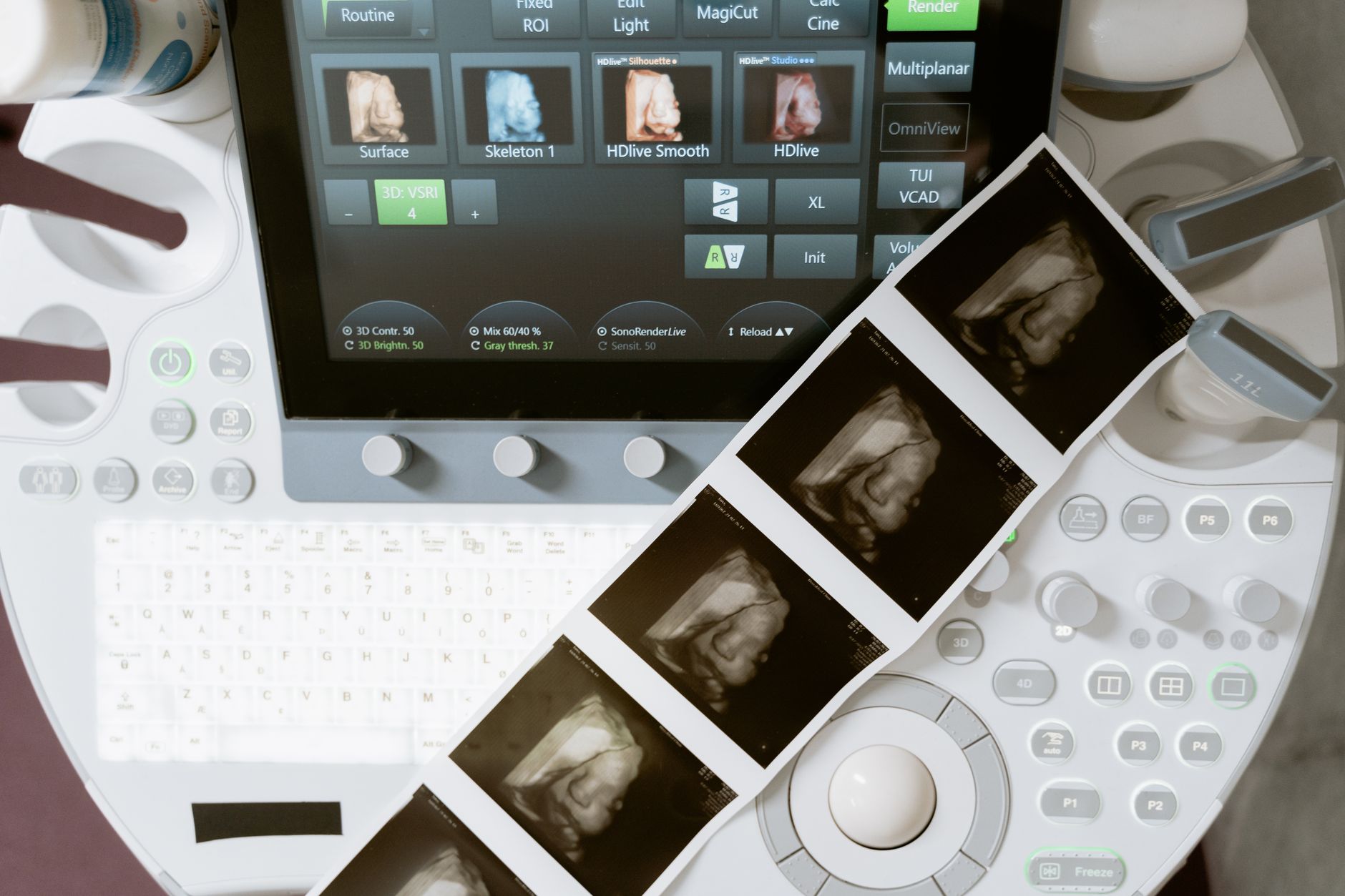
Sonography for pregnant women is scheduled at specific weeks to monitor fetal development; learn the timing of each ultrasound and what it reveals.
By Shubhra Mishra — a mom of two who turned her own confusion during pregnancy into BumpBites, a global mission to make food choices clear, safe, and stress-free for every expecting mother. 💛
Check whether any food is safe during pregnancy with the BumpBites Food Safety Checker.
Quick take: Sonography for pregnant women is safe when performed by qualified providers at the medically indicated times. Typical scans include a 12‑week check‑up, a detailed 20‑week anatomy scan, and later growth scans around 30 weeks. Costs vary, insurance often helps, and you can usually watch the images live online. If you notice severe bleeding, severe pain, or loss of fetal movement, call your care team right away.
It’s 2 a.m., you’re curled up on the couch, and the faint hum of the ultrasound machine from yesterday’s appointment is still in your mind. “Did I do something wrong?” you wonder, scrolling through endless articles about “sonography for pregnant women.” The good news is that most scans are routine, painless, and designed to reassure you and your provider about your baby’s development.
In this guide we walk through every standard ultrasound you’ll encounter, what each one visualizes, how to prepare, safety basics, cost considerations, and what to expect if you have a high‑risk pregnancy. We’ll also answer the most common “what‑if” questions—like spotting twins early or wondering whether an MRI is ever needed.
By the end you’ll know exactly when each scan happens, what the images mean, and how to talk confidently with your obstetric team.
What does a 12‑week ultrasound show for pregnant women?
A 12‑week sonogram, often called the “dating scan,” is usually the first detailed look at your baby. At this stage the embryo is about 5 cm long and the following structures can be seen:
Heart activity: A steady “ticky‑tack” beat confirms viability.
Crown‑rump length (CRL): The measurement from the top of the head to the buttocks helps calculate gestational age.
Gestational sac and yolk sac: These support structures confirm a healthy early pregnancy.
Number of embryos: The scan can reveal twins or higher‑order multiples, even if you haven’t heard the news yet.
Basic anatomy: The limb buds, nasal bone, and early brain structures become visible.
Most providers use this scan to verify the due date and rule out early complications such as ectopic pregnancy. A clear heartbeat at 12 weeks is reassuring, and any major abnormality is rare at this point, though subtle issues may be noted for follow‑up.
The 12‑week scan often reveals the first heartbeat and basic anatomy.
When is the first sonography appointment recommended during pregnancy?
The first sonography appointment is typically scheduled between 6 and 10 weeks gestation, depending on your provider’s protocol and whether you have any risk factors. This early scan is usually a transvaginal ultrasound, which offers clearer images of the gestational sac and can confirm an intrauterine pregnancy.
If you have a history of miscarriage, an irregular menstrual cycle, or are undergoing fertility treatment, your doctor may schedule the first scan sooner—sometimes as early as 5 weeks—to ensure the pregnancy is developing in the uterus and not elsewhere.
After the initial check, the next standard scan is the 12‑week dating scan described above, followed by the 20‑week anatomy scan. High‑risk patients may have additional early scans to monitor growth, placenta position, or Doppler blood flow.
How accurate is the anatomy scan at 20 weeks?
The 20‑week anatomy scan, also called the “mid‑trimester scan,” is the most comprehensive routine ultrasound. According to the American College of Obstetricians and Gynecologists (ACOG), it detects major structural anomalies with a sensitivity of roughly 95 % for conditions such as neural tube defects, major heart defects, and abdominal wall defects.
During this scan the sonographer measures head circumference, abdominal circumference, femur length, and other biometric data to assess growth. The placenta’s position, the amount of amniotic fluid, and the baby’s spine and limbs are also evaluated.
While the scan is highly accurate for major defects, it may miss subtle abnormalities that become apparent later in pregnancy or after birth. If a concern arises, a follow‑up targeted ultrasound or fetal MRI may be recommended.
What can a 30‑week ultrasound reveal about baby’s growth?
By the third trimester, the baby’s size and position become the focus. A 30‑week (or 28‑32 week) growth scan typically includes:
Biometry: Head circumference, abdominal circumference, and femur length are measured to confirm that the baby is growing at the expected rate.
Amniotic fluid volume: Too much or too little fluid can signal complications.
Placenta location and maturity: The sonographer checks for signs of placental insufficiency.
Fetal position: Determining whether the baby is breech or head‑down helps plan delivery.
Estimated weight: While not exact, this estimate helps assess whether the baby is small‑for‑gestational‑age (SGA) or large‑for‑gestational‑age (LGA).
For parents, this scan often provides the first clear view of the baby’s face, limbs, and movements—an emotional milestone that many families cherish.
The 30‑week scan helps assess growth, fluid levels, and baby’s position.
Differences between a routine prenatal ultrasound and a high‑risk sonogram
Routine prenatal ultrasounds follow a standard schedule (12 weeks, 20 weeks, 28‑32 weeks, and sometimes 36 weeks) and focus on confirming anatomy and growth. High‑risk sonograms are tailored to specific concerns and may include:
More frequent scans: Women with hypertension, diabetes, or previous preterm birth may have weekly or bi‑weekly Doppler studies.
Specialized Doppler assessments: Measuring blood flow in the umbilical artery, ductus venosus, and middle cerebral artery to evaluate fetal well‑being.
Targeted anomaly scans: If a concern such as a possible heart defect is raised, a focused exam with higher resolution is performed.
Extended imaging time: Sessions may last longer to capture detailed measurements.
High‑risk scans are usually performed by a maternal‑fetal medicine specialist or a sonographer with advanced training. The goal is early detection of issues that could affect the pregnancy’s outcome, allowing timely intervention.
Is it normal to see twins on a 14‑week ultrasound?
Yes. By 14 weeks the two embryos are usually large enough to be distinguished as separate heartbeats, sacs, and bodies. Many parents discover they are expecting twins (or higher multiples) during the 12‑ to 14‑week scan, even if they hadn’t known before.
Seeing twins early is reassuring because it gives your care team time to monitor growth, plan appropriate nutrition, and schedule additional scans. However, twins also carry higher risks for preterm birth, gestational diabetes, and preeclampsia, so your provider will likely recommend more frequent monitoring.
What are the signs of placenta previa on an ultrasound?
Placenta previa occurs when the placenta partially or completely covers the cervical opening. Ultrasound is the primary tool for diagnosing it. Typical sonographic signs include:
Low‑lying placenta: The placenta is seen within 2 cm of the internal cervical os.
Complete coverage: The placenta fully overlaps the os, classified as complete previa.
Partial coverage: The placenta covers part of the os, known as marginal or partial previa.
Most cases are identified in the second trimester, and many resolve as the uterus expands. Your provider will follow up with repeat scans around 28 weeks to see if the placenta has moved away from the cervix.
What is the cost of prenatal ultrasound in the US?
Out‑of‑pocket costs for a standard prenatal ultrasound in the United States typically range from $150 to $300 per scan. Prices vary based on:
Geographic location (urban centers often cost more).
Facility type (hospital vs. independent imaging center).
Whether the scan is considered routine or diagnostic.
Insurance coverage and deductible status.
Scan type
Typical out‑of‑pocket cost
Insurance coverage*
12‑week dating scan
$150–$250
Usually covered 80–100 %
20‑week anatomy scan
$200–$300
Often fully covered
High‑risk Doppler studies
$250–$400
Coverage varies; prior authorization common
*Coverage depends on your plan’s network status and whether a physician’s order is on file. Always check with your insurer before scheduling.
How to prepare for a fetal ultrasound appointment
Preparation is simple but makes a big difference in image quality:
Full bladder: Drink 16–20 oz of water 30–45 minutes before the appointment. A full bladder pushes the uterus into a better viewing position.
Comfortable clothing: Wear loose‑fitting tops or a shirt that can be easily lifted or a maternity dress.
Timing: Early‑morning appointments are ideal for a full bladder without discomfort.
Bring your records: If you have prior scans, bring the reports so the sonographer can compare.
Relax: Deep breathing helps you stay still, which improves image clarity.
For later‑stage scans, a full bladder is less critical, but you may still be asked to drink a small amount to help visualize the placenta.
Can I watch my baby’s ultrasound live online?
Many modern clinics offer live streaming options for a small fee. The video feed is typically displayed on a tablet or computer in the waiting room, and some providers send a secure link that lets you view the images in real time from your phone.
Live streaming does not affect the safety or quality of the scan. However, keep in mind that the primary goal of the sonographer is to obtain accurate measurements, so it’s best to stay focused on the experience rather than trying to capture every moment on video.
Ultrasound safety guidelines for pregnant women
Ultrasound uses non‑ionizing sound waves, which are considered safe when applied according to the “ALARA” principle—As Low As Reasonably Achievable. The U.S. Food and Drug Administration (FDA) and the American Institute of Ultrasound in Medicine (AIUM) recommend the following safeguards:
Use the lowest possible acoustic output that still yields a clear image.
Limit the duration of each scan to the time needed for the clinical question (usually 20–30 minutes).
Avoid “keeps‑alive” scans that are performed solely for entertainment without a medical indication.
Extensive research, including large cohort studies reviewed by ACOG, has found no evidence of harmful effects on the fetus from routine prenatal ultrasounds. Nonetheless, if you have concerns, discuss them with your provider.
What does a Doppler ultrasound measure in pregnancy?
Doppler ultrasound evaluates blood flow within fetal vessels and the placenta. The most common Doppler measurements include:
Umbilical artery flow: Assesses resistance; high resistance may signal placental insufficiency.
Middle cerebral artery (MCA) flow: Detects fetal anemia or hypoxia.
Ductus venosus flow: Provides insight into cardiac function.
These measurements are especially valuable for pregnancies complicated by diabetes, hypertension, or growth restriction. Results guide decisions about timing of delivery or interventions like steroids for lung maturity.
How many ultrasounds are safe during pregnancy?
There is no fixed “maximum” number of ultrasounds, but ACOG advises that each scan be medically justified. Routine care typically includes 4–5 scans, while high‑risk pregnancies may require additional targeted studies. The key is that the ultrasound’s benefit outweighs any theoretical risk.
In practice, most obstetricians follow a schedule that aligns with the standard timeline (first‑trimester dating, 20‑week anatomy, 28‑32‑week growth, and a final near‑term scan). If your provider recommends extra scans, they will explain the specific reason—such as monitoring a low‑lying placenta or fetal growth.
Ultrasound vs MRI for fetal imaging
Ultrasound remains the first‑line imaging tool because it is inexpensive, widely available, and safe. Magnetic resonance imaging (MRI) is reserved for situations where ultrasound cannot provide sufficient detail, such as:
Complex brain anomalies that need high‑resolution imaging.
Suspected spinal cord defects.
Evaluation of the fetal thorax when ultrasound is limited by maternal habitus.
MRI does not use ionizing radiation, but it is more costly and usually performed after the second trimester. According to the National Institute for Health and Care Excellence (NICE), MRI is recommended only after a targeted ultrasound raises a specific concern.
From our medical team: “All standard prenatal ultrasounds are safe when performed by certified professionals. If you have a high‑risk condition, the extra scans we recommend are designed to monitor your baby’s health closely. Always feel free to ask your provider why a particular scan is needed, and never hesitate to discuss any worries about safety or cost.”
Myth vs. fact
Myth: “You can have as many ultrasounds as you want for fun.”
Fact: While ultrasounds are safe, they should be performed only when medically indicated. Unnecessary “keeps‑alive” scans add cost and may expose the fetus to higher acoustic output without benefit.
Myth: “Ultrasound can determine the baby’s exact gender at 12 weeks.”
Fact: Gender can often be seen after 14 weeks, but the accuracy at 12 weeks is limited. Most providers wait until the 20‑week anatomy scan for reliable gender identification.
Myth: “If an ultrasound looks normal, no further testing is needed.”
Fact: Normal findings at one stage do not guarantee the absence of later‑developing issues. Ongoing monitoring follows the standard schedule, especially for high‑risk pregnancies.
Key takeaways
Standard sonography for pregnant women includes a 12‑week dating scan, a 20‑week anatomy scan, and later growth scans around 28‑32 weeks and 36 weeks.
Ultrasound is safe when performed by qualified providers using the ALARA principle; no proven harm exists for routine scans.
High‑risk pregnancies may need extra Doppler studies, more frequent scans, and possibly MRI for detailed assessment.
Typical US costs range $150‑$300 per scan; insurance often covers routine scans, but out‑of‑pocket fees vary.
Prepare with a full bladder, comfortable clothing, and a relaxed mindset to get the best images.
If you notice severe abdominal pain, heavy bleeding, loss of fetal movement, or any concerning symptoms, contact your care team immediately.
Frequently asked questions
How often should I get an ultrasound during pregnancy?
Most pregnancies include 4–5 routine ultrasounds (12 weeks, 20 weeks, 28‑32 weeks, and a late‑term scan). High‑risk women may have additional targeted scans as advised by their provider.
Can an ultrasound detect birth defects?
Yes, the 20‑week anatomy scan can identify many major structural anomalies with about 95 % sensitivity. Some subtle defects may be missed and are evaluated later with targeted ultrasound or fetal MRI.
Is it safe to have an ultrasound in the first trimester?
Yes. First‑trimester scans are considered safe when performed by trained professionals. The FDA and ACOG affirm that no harmful effects have been demonstrated for routine early ultrasounds.
What does the baby look like on a 20‑week ultrasound?
At 20 weeks the baby is roughly the size of a banana, with clearly visible arms, legs, facial features, and a beating heart. The anatomy scan provides a detailed view of the brain, spine, and major organs.
Do I need a doctor’s referral for a prenatal ultrasound?
In the United States, most insurers require a physician’s order for coverage. Even if you can self‑pay, an obstetrician’s referral ensures the scan is timed appropriately and interpreted correctly.
Can an ultrasound determine the baby’s gender?
Gender can usually be identified after 14 weeks, with the most reliable determination at the 20‑week anatomy scan. Accuracy before this window is limited.
When to call your doctor
If you experience any of the following, seek medical attention right away: heavy vaginal bleeding, severe abdominal pain, sudden loss of fetal movement, fever with chills, or a sudden change in your baby’s heartbeat pattern as heard on a home Doppler device.
This article is for informational purposes only and does not replace personalized medical advice. Always discuss your specific situation with your obstetric provider.
References
American College of Obstetricians and Gynecologists (ACOG). “Ultrasound Guidance for Prenatal Care,” Practice Bulletin No. 183, 2022.
U.S. Food and Drug Administration (FDA). “Diagnostic Ultrasound Safety,” 2021.
American Institute of Ultrasound in Medicine (AIUM). “Guidelines for the Safe Use of Ultrasound in Pregnancy,” 2020.
National Institute for Health and Care Excellence (NICE). “Fetal Anomaly Screening,” NG152, 2021.
Society for Maternal-Fetal Medicine (SMFM). “Guidelines for the Use of Doppler Ultrasound in Pregnancy,” 2023.
Centers for Disease Control and Prevention (CDC). “Prenatal Care and Imaging,” 2022.
World Health Organization (WHO). “Recommendations on Antenatal Care for a Positive Pregnancy Experience,” 2016.
Mayo Clinic. “Ultrasound during pregnancy: What to expect,” 2023.
National Health Service (NHS). “Ultrasound scans in pregnancy,” 2022.
Not sure about the label on Pregnancy Ultrasound Guide When You Get Each One What They Show products?
Snap the ingredients list and SafeFilter checks every ingredient for your stage — only 3 free scans this month, then you're locked until reset. Unlimited from $7/mo or $50/yr.
When Shubhra Mishra was expecting her first child in 2016, she was overwhelmed by conflicting food advice — one site said yes, another said never. By the time her second baby arrived in 2019, she realized millions of mothers face the same confusion.
That sparked a five-year journey through clinical nutrition papers, cultural diets, and expert conversations — all leading to BumpBites: a calm, compassionate space where science meets everyday motherhood.
Her long-term vision is to build a global community ensuring safe, supported, and free deliveriesfor every mother — because no woman should face pregnancy alone or uninformed. 🌿
🌍 Stand with mothers, shape safer guidance
Join a small circle of experts who review BumpBites articles so expecting parents everywhere can decide with confidence.