Most pregnancies require 1-3 ultrasounds. Learn how many ultrasounds during pregnancy are recommended, when they’re done, and why they’re essential for your baby’s health.
By Shubhra Mishra — a mom of two who turned her own confusion during pregnancy into BumpBites, a global mission to make food choices clear, safe, and stress-free for every expecting mother. 💛
Check whether any food is safe during pregnancy with the BumpBites Food Safety Checker.
Download the Complete Pregnancy Food Guide (10,000 Foods) 📘
Instant PDF download • No spam • Trusted by thousands of moms
💡 Your email is 100% safe — no spam ever.
Quick take: Most pregnancies involve 2 to 3 routine ultrasounds, but the exact number depends on your health, the trimester, and whether you have a high‑risk pregnancy. Ultrasound is a safe, non‑invasive way to track your baby’s growth and spot potential concerns; there’s no evidence that medically‑recommended scans harm the developing fetus.
It’s 2 a.m., you’re curled up on the couch, and a sudden flutter makes you wonder: “How many ultrasounds will I need before my baby arrives?” You’ve probably heard a mix of stories—some moms say they had five scans, others only one. The good news is that the answer isn’t one‑size‑fits‑all. In this guide we’ll break down what the standard schedule looks like, why doctors recommend extra scans for certain conditions, and how you can prepare for each appointment.
We’ll walk through the typical ultrasound timeline, explain what you’ll see at the 20‑week scan, discuss the safety of repeated imaging, and give you practical tips for each trimester. By the end you’ll have a clear picture of what’s normal, what’s optional, and when you might need a little extra reassurance.
How many ultrasounds during a normal pregnancy?
For most low‑risk pregnancies, guidelines from the American College of Obstetricians and Gynecologists (ACOG) and the UK’s National Institute for Health and Care Excellence (NICE) recommend at least two routine ultrasounds: one in the first trimester (often called the dating scan) and a detailed anatomy scan around 18‑20 weeks. Some clinicians add a third scan in the third trimester to check growth and amniotic fluid levels, especially if you’re over 35 or have a higher BMI.
The average number of ultrasounds reported in large U.S. cohort studies is 2.4 per pregnancy. In the United Kingdom, the NHS typically schedules two scans for uncomplicated pregnancies, with a third offered if there are concerns about fetal growth or position. In practice, most women end up having 2 to 3 scans, and a handful may have 4 or 5 if additional monitoring is needed.
Why the variation? Ultrasound is both a diagnostic tool and a reassurance for parents. The first‑trimester scan confirms the due date, checks for a single heartbeat, and can catch early issues like ectopic pregnancy. The mid‑pregnancy anatomy scan evaluates the baby’s organs, measures growth, and often reveals the baby’s sex—information that can guide later care decisions. A third‑trimester scan assesses growth trajectory, placenta location, and fluid levels, helping the team decide on timing of delivery.
When you’re planning your schedule, ask your provider: “Based on my health and my baby’s growth, how many scans do you anticipate?” This question sets the stage for a personalized plan that aligns with both evidence‑based guidelines and your peace of mind.
It’s also worth noting that many clinics now bundle the dating and early anatomy scans into a single appointment if your schedule is tight, which can reduce the total number of visits without sacrificing information. If you’re traveling long distances for care, discuss the possibility of combined appointments early on.
Typical first‑trimester ultrasound: confirming dates and heartbeat.
What to expect during an ultrasound at 20 weeks?
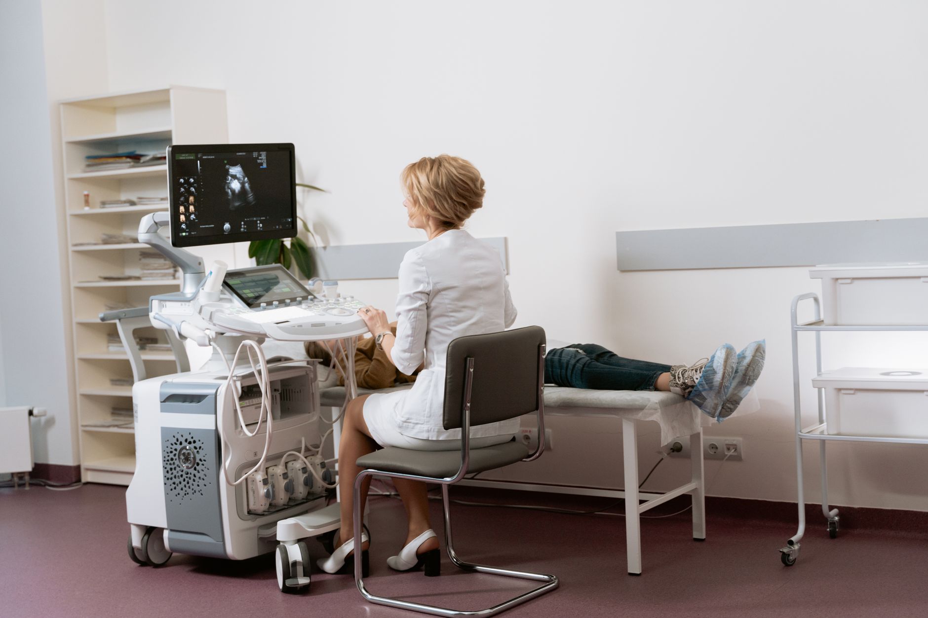
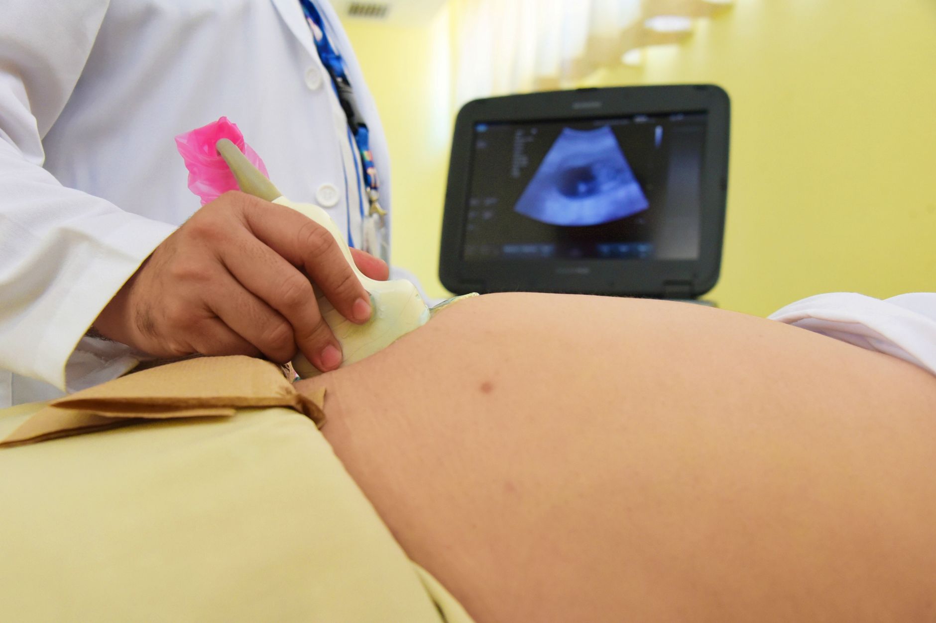
The 20‑week scan, also called the anatomy or level‑II ultrasound, is often the most detailed imaging you’ll have. By this point the baby is about 5 inches long and many organs are fully formed, so the sonographer can assess the heart, brain, kidneys, spine, and even the placenta. You’ll likely hear the baby’s heartbeat twice—once from the transducer on your abdomen and once from a Doppler probe that captures the sound more clearly.
During the exam, the sonographer will apply a warm gel to your abdomen and move a small handheld transducer across your belly. The gel helps conduct sound waves, and the device sends high‑frequency sound that reflects off the baby and surrounding tissues. The reflected echoes create the black‑and‑white image you’ll see on the monitor. It’s normal to feel a slight coolness from the gel and a gentle pressure as the transducer glides.
Typical findings at 20 weeks include:
Fetal measurements: head circumference, abdominal circumference, and femur length to estimate growth.
Organ assessment: visualizing the brain ventricles, heart chambers, kidneys, and stomach.
Placenta location: confirming it’s not covering the cervix (placenta previa).
Amniotic fluid volume: ensuring there’s enough fluid surrounding the baby.
If the sonographer spots anything atypical—like a mild ventriculomegaly or a low‑lying placenta—they’ll note it and discuss whether further testing (such as a targeted ultrasound or MRI) is needed. Most findings are minor and resolve on their own, but the scan offers a crucial checkpoint for early intervention if needed.
Because the 20‑week scan is so comprehensive, many providers will also answer common parental questions on the spot—like “What’s my baby’s sex?” or “Can we see a heartbeat?” If you have specific concerns (e.g., a family history of a particular condition), let the sonographer know before the exam so they can focus on the relevant structures.
How often should I get an ultrasound during pregnancy?
The frequency of ultrasounds depends on three main factors: your overall health, any pregnancy‑related complications, and the stage of gestation. Below is a concise guide:
Trimester
Typical Scan Frequency (Low‑Risk)
Typical Scan Frequency (High‑Risk)
First (0‑13 weeks)
1 (dating scan)
1‑2 (dating + early anomaly if indicated)
Second (14‑27 weeks)
1 (20‑week anatomy)
2‑3 (anatomy + growth + targeted scans)
Third (28‑40 weeks)
0‑1 (growth check if indicated)
1‑2 (growth, placenta, and biophysical profile)
In a standard pregnancy, you’ll likely have three scans: the first‑trimester dating scan, the 20‑week anatomy scan, and a late‑third‑trimester growth scan if your provider feels it’s necessary. High‑risk pregnancies—such as those involving gestational diabetes, preeclampsia, multiple gestations, or a history of fetal growth restriction—often require additional ultrasounds to monitor the baby’s development more closely.
Ask yourself: “Do I have any conditions that could affect fetal growth?” If the answer is yes, discuss a tailored schedule with your obstetrician. The goal is to balance adequate monitoring with the principle of “as low as reasonably achievable” (ALARA) when it comes to any medical imaging.
One practical tip is to keep a calendar (paper or digital) that marks each scheduled ultrasound. Adding a reminder to bring any previous scan reports to the next appointment can streamline discussions and reduce the need for repeat imaging.
Are too many ultrasounds during pregnancy bad for the baby?
Current evidence from ACOG, the FDA, and the World Health Organization (WHO) indicates that diagnostic ultrasound, when performed by a qualified professional at the recommended settings, does not increase the risk of birth defects or developmental problems. The primary safety principle is to keep the acoustic output low and limit exposure time—exactly what “standard” obstetric scans do.
Studies that have examined large populations (more than 200,000 pregnancies) found no link between the number of routine scans and adverse outcomes. However, the guidance cautions against non‑medical “keepsake” ultrasounds that may use higher‑intensity settings or last longer than necessary. If you’re considering an extra scan for a photo or a curiosity, ask your provider whether it meets the ALARA principle and whether it’s medically justified.
In short, too many ultrasounds are not harmful when each scan follows clinical guidelines. The risk only becomes a concern if scans are performed without medical indication, at higher intensities, or for prolonged periods. Your care team will always weigh the benefit of additional imaging against any theoretical risk, keeping your baby’s safety front‑and‑center.
For families who are especially anxious, many clinics now offer “virtual” 3‑D reconstructions that can be shared as keepsake images without requiring an extra scan. These are generated from the data captured during a standard anatomy scan, so they don’t add additional exposure.
How many ultrasounds are needed for a high‑risk pregnancy?
High‑risk pregnancies—such as those involving twins, maternal hypertension, pre‑existing diabetes, or a previous stillbirth—often call for a more intensive ultrasound schedule. A typical high‑risk protocol might include:
Early dating scan (8‑12 weeks): precise gestational age and early anatomy.
First‑trimester anatomy (around 12‑13 weeks): to detect major anomalies early.
Mid‑pregnancy anatomy (18‑20 weeks): detailed organ assessment.
Growth scans (every 4 weeks after 28 weeks): monitoring weight, amniotic fluid, and placental position.
Biophysical profile (BPP) or Doppler studies (as indicated): especially for cases of preeclampsia or fetal growth restriction.
Depending on your situation, you might have 5 to 8 scans total. For example, a mother carrying twins may have an additional scan at 24 weeks to assess twin‑specific growth patterns, and another at 32 weeks to monitor the shared placenta.
It’s helpful to keep a simple tracking sheet: note the date, gestational age, purpose of the scan, and any follow‑up recommendations. This record can make conversations with your obstetrician smoother and ensure no recommended scan is missed.
When you’re in a high‑risk category, your provider may also schedule “targeted” ultrasounds after a concerning lab result (like an abnormal blood pressure reading) to quickly assess whether the baby’s growth is being affected.
Can I have too many ultrasounds during pregnancy?
“Too many” is a relative term. From a medical standpoint, the answer is no—provided each scan is ordered for a specific clinical reason. The concern arises when patients request extra “keepsake” scans that are not medically indicated. Such scans may use higher acoustic power or longer exposure times, which the guidelines advise against for routine use.
If you feel anxious and want reassurance, discuss it openly with your provider. Often, a single extra scan can be scheduled if it’s done with the same low‑output settings used for diagnostic imaging. Many clinics also offer “virtual” 3‑D images that can be shared without additional exposure, as they are derived from the same data set captured during a standard scan.
In practice, most women who have a high‑risk condition will have the recommended number of scans without exceeding safety thresholds. If you ever wonder whether a particular scan is necessary, ask: “What will this scan tell us that we don’t already know?” A clear answer will help you weigh the benefit against any theoretical risk.
Remember that anxiety itself can affect how you perceive the need for imaging. Mind‑body techniques—such as guided breathing or short walks—can reduce the urge for unnecessary reassurance scans while you await scheduled appointments.
Ultrasound schedule during pregnancy by trimester
Below is a trimester‑by‑trimester overview that aligns with both ACOG and NICE guidelines, while also addressing common patient questions.
First Trimester (0‑13 weeks): A dating scan at 8‑12 weeks confirms gestational age, checks for a single heartbeat, and can detect early ectopic pregnancy. Some providers add a nuchal translucency (NT) scan if you’re undergoing first‑trimester screening for chromosomal anomalies.
Second Trimester (14‑27 weeks): The anatomy scan at 18‑20 weeks evaluates organ development, measures growth, and assesses placental location. A follow‑up scan at 24‑28 weeks may be ordered if there are concerns about growth or if you have gestational diabetes.
Third Trimester (28‑40 weeks): Growth scans every 4 weeks (or more frequently if indicated) monitor fetal size, amniotic fluid volume, and placental health. A biophysical profile (BPP) may be performed if you develop preeclampsia or decreased fetal movements.
For low‑risk pregnancies, a typical schedule looks like this:
8‑12 weeks: dating scan
18‑20 weeks: anatomy scan
32‑36 weeks (optional): growth check
High‑risk pregnancies may add scans at 24, 28, 32, 36 weeks, and possibly a targeted scan for specific concerns (e.g., placental insufficiency). Your provider will customize the timetable based on your health history and any emerging findings.
When you receive a scan report, ask your provider to highlight any measurements that fall outside the typical range. Understanding the numbers (like estimated fetal weight or amniotic fluid index) can demystify the process and reduce the need for extra reassurance scans.
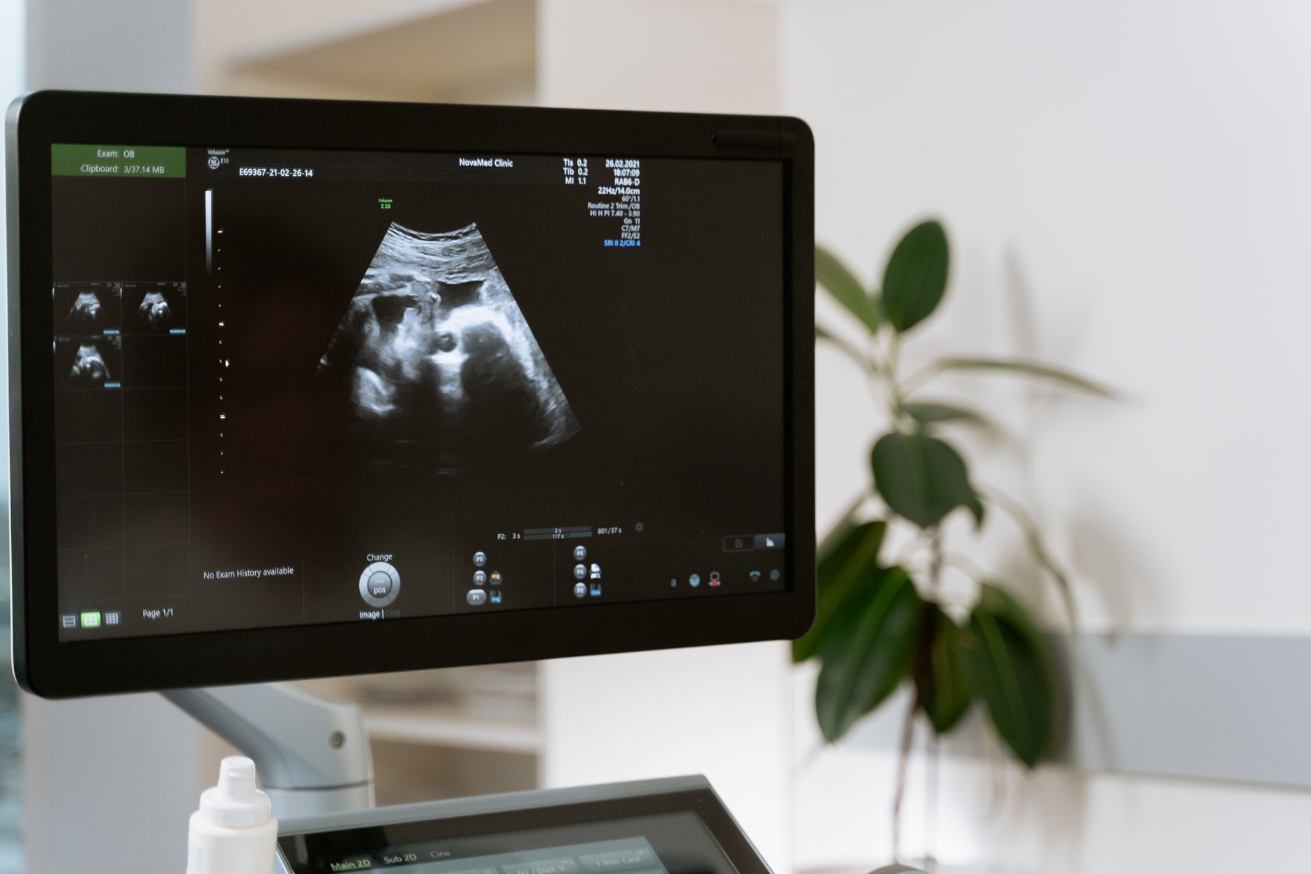
Typical ultrasound timeline for a low‑risk pregnancy.
Why are multiple ultrasounds necessary during pregnancy?
Multiple ultrasounds serve three core purposes: monitoring growth, detecting complications early, and providing reassurance. Growth monitoring is essential because fetal size can influence delivery timing. For example, a baby that is significantly larger than the estimated gestational age may increase the risk of shoulder dystocia, while a small‑for‑gestational‑age baby might signal placental insufficiency.
Complication detection is another key reason. Ultrasound can reveal placental previa (placenta covering the cervix), abruption (premature separation), or oligohydramnios (low amniotic fluid). Early identification of these issues allows your care team to intervene—sometimes with medication, increased monitoring, or planned early delivery—to improve outcomes.
Finally, the emotional benefit should not be underestimated. Seeing your baby’s heartbeat and movements on a screen can reduce anxiety and strengthen the parental bond. Many parents cite the 20‑week anatomy scan as a memorable moment that solidifies the reality of pregnancy.
In short, each scan adds a layer of information that helps clinicians tailor care to your baby’s unique trajectory, while also giving you a window into the miracle growing inside you.
From our medical team: Ultrasound is a cornerstone of prenatal care because it provides real‑time, radiation‑free imaging. If you ever feel uncertain about the number of scans you’ve been recommended, bring a written summary of the schedule to your next appointment. This makes it easier for you and your provider to discuss the purpose of each scan, address any safety concerns, and ensure you’re getting the right amount of monitoring for your specific situation.
What are 3‑D and 4‑D ultrasounds, and are they safe?
Three‑dimensional (3‑D) and four‑dimensional (4‑D) ultrasounds create detailed images that can be viewed from multiple angles, and 4‑D adds real‑time motion. They are often marketed as “keepsake” scans because they produce lifelike pictures of the baby’s face or movements. The technology uses the same acoustic principles as standard 2‑D scans, but the transducer collects a larger data set that is processed by advanced software.
Safety-wise, 3‑D/4‑D scans are considered safe when performed by a qualified sonographer using the same low‑output settings as routine scans. The FDA and ACOG emphasize that any ultrasound should adhere to the ALARA principle, so a single 3‑D/4‑D session is unlikely to pose a risk. However, they are not medically necessary for most pregnancies, and repeated “keepsake” sessions could add unnecessary exposure. If you’re interested in a 3‑D image, ask whether the same data can be captured during your scheduled anatomy scan, which often eliminates the need for an extra appointment.
Some parents find that seeing a 3‑D image helps them bond with their baby, especially if they had a difficult conception journey. If you decide to proceed, make sure the clinic documents that the scan was performed with standard safety settings and that the images are stored as part of your medical record.
3‑D ultrasound can produce detailed images of the baby's face, but safety follows the same guidelines as standard scans.
Can I have a home or telehealth ultrasound?
Advances in portable ultrasound technology have made home‑based scans technically possible, but they are not yet a standard part of prenatal care in most health systems. Portable devices require a trained operator to obtain reliable images, and interpretation still needs a qualified sonographer or physician. Some research studies (e.g., a 2022 trial published by the Society for Maternal‑Fetal Medicine) showed that trained midwives using handheld units could acquire adequate images for certain assessments, but widespread adoption is limited by regulatory and reimbursement factors.
Telehealth‑enabled ultrasound works differently: you attend a clinic where a technician performs the scan, and the images are streamed live to a specialist who reviews them remotely. This model can increase access in rural areas and reduce travel time, while still maintaining the safety and quality standards of in‑person imaging. If you live far from a hospital, ask whether your provider offers a tele‑ultrasound service or a partnership with a nearby imaging center.
At this time, most insurance plans (including Medicaid and many private carriers) only cover scans performed in accredited facilities. Home scans that are not medically indicated are typically out‑of‑pocket. Always confirm coverage before scheduling a non‑standard appointment.
Telehealth‑enabled ultrasounds can bring specialist review to a local clinic, improving access without compromising safety.
Myth vs. fact
Myth: “All pregnant women should have at least five ultrasounds.”
Fact: The standard recommendation for low‑risk pregnancies is two to three scans; more are only ordered when clinically indicated.
Myth: “Ultrasound can cause birth defects if done too often.”
Fact: Diagnostic ultrasound, performed with proper settings, has not been shown to increase the risk of birth defects. The safety guidelines focus on limiting unnecessary exposure.
Myth: “If I miss a scheduled scan, my baby’s health is jeopardized.”
Fact: While regular monitoring is valuable, missing a single routine scan rarely leads to serious complications. Your provider can adjust the schedule if needed.
Key takeaways
Most low‑risk pregnancies have 2 to 3 ultrasounds; high‑risk pregnancies may need 5 or more.
The 20‑week anatomy scan is the most comprehensive and checks organ development, growth, and placenta.
Diagnostic ultrasound is safe when performed by qualified professionals using standard settings.
Extra “keepsake” scans are optional but should follow the ALARA principle to avoid unnecessary exposure.
Keep a simple log of scan dates, gestational ages, and findings to stay organized.
Always discuss any anxiety or questions with your obstetrician; they can tailor the schedule to your needs.
Frequently asked questions
How many ultrasounds are normal during pregnancy?
For a typical low‑risk pregnancy, 2 to 3 ultrasounds—one in the first trimester and one detailed scan at 18‑20 weeks—are considered normal; a third later‑trimester scan may be added if growth concerns arise.
What is the purpose of multiple ultrasounds during pregnancy?
Multiple scans allow clinicians to track fetal growth, identify structural anomalies, monitor placental position, and detect complications such as growth restriction or low amniotic fluid, all while providing reassurance to parents.
Can too many ultrasounds harm the baby?
Current evidence from ACOG and WHO shows that medically indicated ultrasounds, even when repeated, do not increase the risk of birth defects or developmental problems; the risk only exists with non‑medical, high‑intensity scans.
How often should I get an ultrasound during my first trimester?
Most providers recommend a single dating scan between 8 and 12 weeks; if you’re undergoing early screening for chromosomal anomalies, a nuchal translucency scan may be added around 11‑13 weeks.
Are ultrasounds during pregnancy covered by insurance?
In the United States, most private insurers and Medicaid cover the standard prenatal ultrasound schedule (dating and anatomy scans). Coverage for additional scans depends on medical necessity and your plan’s specifics.
What are the risks of not having enough ultrasounds during pregnancy?
Missing recommended scans can delay the detection of growth abnormalities, placental issues, or congenital anomalies, potentially limiting timely interventions; however, occasional gaps can be mitigated by close clinical monitoring and symptom awareness.
Can I request a 3‑D “keepsake” ultrasound?
Yes, many clinics offer 3‑D/4‑D scans as an optional service, but they are not medically necessary; if you choose one, ask that it be performed with the same low‑output settings used for routine imaging to stay within safety guidelines.
Is a telehealth ultrasound as accurate as an in‑person scan?
When a qualified sonographer performs the scan and a specialist reviews the images remotely, tele‑ultrasound can be just as accurate for routine assessments, though insurance coverage may vary and some complex evaluations still require an on‑site visit.
When to call your doctor
If you notice any of the following after an ultrasound—or at any time—contact your obstetrician or midwife promptly:
Decreased fetal movements or a sudden change in activity level.
Bleeding, cramping, or severe abdominal pain.
Persistent fever, chills, or flu‑like symptoms.
Any new concern about the ultrasound findings that you don’t understand.
These signs may indicate a complication that requires urgent evaluation. This article is for informational purposes only and does not replace personalized medical advice.
References
American College of Obstetricians and Gynecologists. “Ultrasound in Pregnancy.” ACOG Committee Opinion No. 730, 2023.
National Institute for Health and Care Excellence. “Routine Antenatal Care.” NICE guideline NG121, 2022.
World Health Organization. “Recommendations on Antenatal Care for a Positive Pregnancy Experience.” WHO, 2021.
U.S. Food and Drug Administration. “Ultrasound Devices: Safety and Efficacy.” FDA Guidance, 2022.
Society for Maternal–Fetal Medicine. “Guidelines for the Use of Ultrasound in High‑Risk Pregnancies.” SMFM Clinical Consensus, 2023.
British Medical Ultrasound Society. “Diagnostic Ultrasound Safety.” BMUS Position Statement, 2022.
Centers for Disease Control and Prevention. “Prenatal Care and Ultrasound Use.” CDC, 2023.
Society for Maternal‑Fetal Medicine. “Handheld Ultrasound in Rural Settings: A 2022 Clinical Trial.” SMFM Journal, 2022.
American College of Radiology. “ALARA Principle in Obstetric Imaging.” ACR White Paper, 2021.
Editor's pick for this topic
About the Author
When Shubhra Mishra was expecting her first child in 2016, she was overwhelmed by conflicting food advice — one site said yes, another said never. By the time her second baby arrived in 2019, she realized millions of mothers face the same confusion.
That sparked a five-year journey through clinical nutrition papers, cultural diets, and expert conversations — all leading to BumpBites: a calm, compassionate space where science meets everyday motherhood.
Her long-term vision is to build a global community ensuring safe, supported, and free deliveriesfor every mother — because no woman should face pregnancy alone or uninformed. 🌿
🌍 Stand with mothers, shape safer guidance
Join a small circle of experts who review BumpBites articles so expecting parents everywhere can decide with confidence.