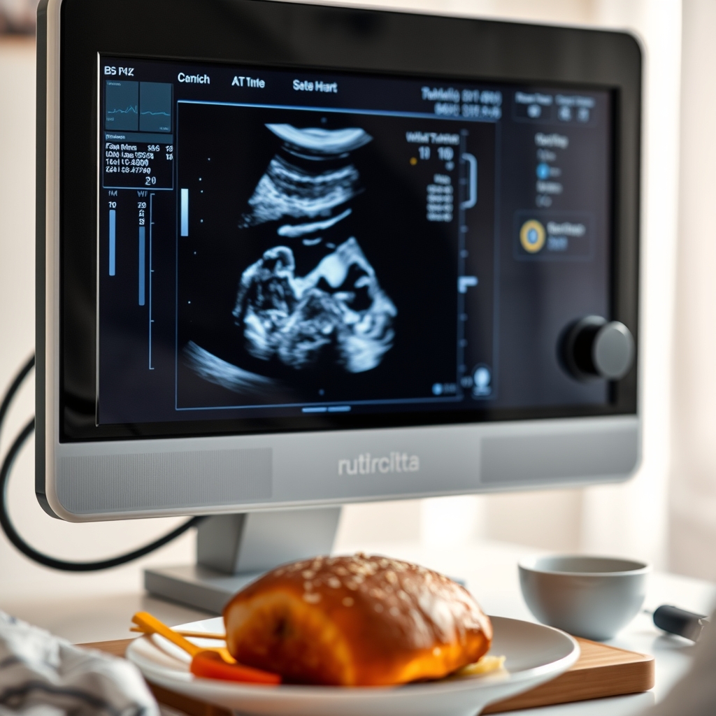
A fetal echocardiogram is needed if risk factors like family history, maternal conditions, or abnormal ultrasounds are present. Learn when this test is essential for your baby’s heart health.
By Shubhra Mishra — a mom of two who turned her own confusion during pregnancy into BumpBites, a global mission to make food choices clear, safe, and stress-free for every expecting mother. 💛
Check whether any food is safe during pregnancy with the BumpBites Food Safety Checker.
Quick take: A fetal echocardiogram is a specialized ultrasound that looks at your baby’s heart. It’s usually recommended when risk factors or concerning signs are present, can be safely done as early as 12–14 weeks, and is non‑invasive for both mother and baby. Most insurers cover it when it’s medically indicated; otherwise costs vary.
It’s 2 a.m., you’re scrolling through a list of “what‑if” questions, and you stumble on the term “fetal echocardiogram.” Your heart races—what does it mean, and do you need one? You’re not alone. Many expectant parents wonder whether this extra test is necessary, safe, or affordable. In this guide we’ll walk through everything you need to know: when doctors recommend it, what signs prompt the test, how early it can be performed, what the procedure looks like, which heart conditions it can reveal, and how insurance and costs play into the picture.
By the end of this article you’ll have a clear picture of the role this imaging plays in prenatal care, what to expect at your appointment, and how to talk with your provider about next steps. Let’s demystify the fetal echocardiogram together, one question at a time.
Seeing your baby’s heart in real time can ease anxiety and guide care.
When is a fetal echocardiogram recommended during pregnancy?
Across the United States and the United Kingdom, obstetric guidelines from the American College of Obstetricians and Gynecologists (ACOG) and the Royal College of Obstetricians and Gynaecologists (RCOG) agree that a fetal echocardiogram is typically advised when there’s an elevated risk of congenital heart disease (CHD). Common scenarios include:
Maternal conditions such as diabetes (especially if poorly controlled), lupus, or a history of heart disease.
A family history of CHD or a known genetic syndrome (e.g., Down syndrome, Turner syndrome).
Abnormal findings on a standard anatomy sweep—like a deviated heart position, abnormal blood flow, or a single‑ventricle appearance.
Exposure to teratogenic medications (e.g., isotretinoin) or significant environmental toxins.
Maternal infections like rubella or cytomegalovirus during the first trimester.
In practice, your obstetrician will schedule the scan when any of these risk factors are present, usually between 18 and 24 weeks gestation. Early detection allows for detailed counseling, possible in‑utero interventions, and birth planning at a center equipped for neonatal cardiac care.
Guidelines from the National Institute for Health and Care Excellence (NICE) also emphasize that any ultrasound anomaly suggestive of cardiac pathology—such as a thickened pericardial effusion—should trigger a referral. The aim is to give families enough time to consider options, from fetal therapy to delivery at a tertiary cardiac unit.
What are the signs that indicate need for a fetal echocardiogram?
Even if you have no known risk factors, certain ultrasound findings can raise a red flag. The most common “signs” that prompt a referral include:
Abnormal cardiac axis: The heart is positioned more to the left or right than usual.
Irregular heart rhythm: The fetus shows tachycardia (fast heart rate) or bradycardia (slow heart rate) on Doppler.
Fluid accumulation: Excess fluid around the lungs (hydrops) may suggest cardiac strain.
Structural anomalies: A missing or malformed atrial or ventricular septum seen on the routine anatomy scan.
Abnormal blood flow patterns: Turbulent flow across the heart chambers or great vessels.
When any of these are spotted, the sonographer will often recommend a dedicated fetal echocardiogram to get a clearer view. In many cases, the “sign” is subtle—a slight deviation that only an experienced fetal cardiologist can interpret.
In addition to the classic ultrasound markers, some labs now incorporate maternal serum biomarkers (e.g., elevated PAPP‑A) into risk stratification. While not diagnostic, these labs can tip the balance toward ordering a fetal echocardiogram when combined with imaging concerns.
How early can a fetal echocardiogram be performed?
Advances in ultrasound technology mean that a fetal echocardiogram can be attempted as early as 12–14 weeks, though most centers prefer waiting until 18–20 weeks when the heart has developed enough detail for accurate assessment. Early scans are technically possible but may have limited resolution, leading to false‑negative results.
For families with a strong suspicion of a cardiac anomaly—such as a known genetic condition—a 12‑week scan can provide an initial glimpse, allowing for a follow‑up later in the second trimester. However, the consensus from ACOG and NICE (National Institute for Health and Care Excellence) is to schedule the definitive fetal echocardiogram between 20 and 24 weeks for the most reliable images.
When an early scan is performed, clinicians often repeat the exam at 20–22 weeks to confirm findings. This two‑step approach balances the desire for early information with the need for diagnostic certainty.
What is the procedure and safety of a fetal echocardiogram?
The fetal echocardiogram is essentially a high‑resolution ultrasound. A trained fetal cardiologist or sonographer places a transducer on your abdomen, and the device emits sound waves that bounce off the baby’s heart, creating detailed images on a monitor. The whole exam typically lasts 30–45 minutes, though the exact time can vary based on fetal position and the complexity of the case.
Because it uses sound waves—not ionizing radiation—it’s considered safe for both mother and baby. The FDA and the American Institute of Ultrasound in Medicine (AIUM) have affirmed that diagnostic ultrasound poses no known risk when performed by qualified professionals. The only precaution is to keep the “thermal index” low, which the equipment automatically regulates. There’s no need for contrast agents or sedation.
After the scan, the technician will review the images and forward them to a fetal cardiologist for interpretation. Most families receive the written report within a week, though some clinics provide a preliminary verbal summary the same day.
For women with pacemakers, implanted neurostimulators, or severe skin sensitivity, the sonographer will take extra care to position the probe to avoid interference, but these situations rarely affect the ability to obtain diagnostic images.
Color Doppler helps visualize blood flow through the tiny chambers.
What conditions can a fetal echocardiogram detect?
Fetal echocardiography is the gold standard for identifying structural heart defects before birth. It can reliably detect:
Congenital heart block associated with maternal antibodies
According to the American Heart Association (AHA), detection rates for major CHD range from 80 % to 95 % when the scan is performed in the optimal window (18–24 weeks) by an experienced fetal cardiologist. Smaller lesions, like tiny muscular VSDs, may be missed but often resolve spontaneously after birth.
Beyond structural anomalies, the exam can also assess functional parameters such as ventricular contractility and myocardial performance index, which can hint at subtle cardiomyopathies that may not become apparent until later in life.
Who pays for a fetal echocardiogram and insurance coverage?
In the United States, most private insurers—including Medicare and Medicaid—cover the test when a physician orders it for a medically indicated reason. The coding typically uses CPT 93325 (fetal echocardiography) and is billed under the maternal health benefit. If you have a high‑deductible plan, you may be responsible for a co‑pay or a portion of the cost.
Without insurance, the out‑of‑pocket price can range from $500 to $1,500, depending on the facility and geographic region. In the United Kingdom, the National Health Service (NHS) provides the scan free of charge when criteria are met; private clinics charge £400–£800.
It’s wise to contact your insurer’s pre‑authorization department early. Ask for a written estimate and verify whether any portion of the cost can be applied to a Flexible Spending Account (FSA) or Health Savings Account (HSA).
Some U.S. states, such as California and New York, have mandates that require insurers to cover fetal echocardiography when a congenital heart defect is suspected, regardless of plan tier. Checking local regulations can prevent unexpected out‑of‑pocket expenses.
Difference between fetal echocardiogram and standard ultrasound
A routine obstetric ultrasound—often called a “growth scan” or “anatomy scan”—captures a broad overview of the baby’s development, placenta, amniotic fluid, and major organs. The sonographer uses a standard transducer and spends a few minutes on each organ system.
By contrast, a fetal echocardiogram is a focused, high‑resolution exam dedicated solely to the heart. It employs specialized probes, color Doppler, and sometimes spectral Doppler to assess blood flow, chamber size, and valve motion. The technician spends significantly more time (30–45 minutes) mapping each cardiac view, and a fetal cardiologist interprets the images for subtle abnormalities.
In short, a standard ultrasound tells you “the baby looks healthy,” while a fetal echocardiogram tells you “the baby’s heart is structurally sound and beating normally.” Both are complementary; the echocardiogram is usually ordered after a routine scan raises a question.
What are the risks of a fetal echocardiogram for mother and baby?
Because the test uses diagnostic ultrasound, the primary risk is minimal. The AIUM and FDA both state that, when performed by qualified personnel, there are no known adverse outcomes for the fetus or mother. The most common “risk” is simply the inconvenience of a longer appointment and the emotional impact of waiting for results.
Rarely, prolonged exposure to high‑intensity ultrasound can cause a temporary rise in tissue temperature, but modern machines automatically limit the output to keep the thermal index below 1.0, well within safety margins. There’s no known link between fetal echocardiography and miscarriage, preterm labor, or developmental issues.
If you have a pacemaker, implanted medical device, or severe skin sensitivity, discuss it with your provider, though it rarely affects the scan.
Fetal echocardiogram cost without insurance
When you pay out‑of‑pocket, expect to spend between $500 and $1,500 in the U.S., depending on the facility’s pricing structure and whether a pediatric cardiologist reads the study. Private hospitals often charge more than community clinics. Some centers offer payment plans or sliding‑scale fees for families without coverage.
Internationally, costs vary widely. In Canada, most provinces cover the test through provincial health plans when indicated, but private clinics can charge CAD 800–CAD 1,200 for a self‑pay appointment.
Average time to get results from a fetal echocardiogram
Most clinics aim to deliver a written report within 7–10 business days. If the scan reveals a possible abnormality, many providers will arrange a follow‑up appointment within 48 hours to discuss findings and next steps. In larger academic centers, preliminary results may be shared on the same day, with the final report arriving a week later.
Some institutions now use secure patient portals that allow you to view the images and preliminary interpretation online within 24 hours, which can reduce anxiety while you wait for the formal report.
Fetal echocardiogram vs newborn echocardiogram
Aspect
Fetal Echocardiogram
Newborn Echocardiogram
Timing
Performed in‑utero, usually 18–24 weeks
Performed after birth, often within the first 24–48 hours
Purpose
Screen for structural defects, plan delivery location
Confirm diagnosis, guide immediate postnatal care
Image clarity
Limited by fetal position, amniotic fluid
Higher resolution, no maternal tissue interference
Risk
Non‑invasive, no radiation
Non‑invasive, safe for newborns
Follow‑up
Potential for in‑utero intervention or delivery planning
Surgical or medical treatment after birth
How to prepare for a fetal echocardiogram appointment
Preparation is straightforward. On the day of the scan:
Wear comfortable, loose‑fitting clothing that allows easy access to your abdomen.
Drink a moderate amount of water (about 2 cups) an hour before the appointment; a full bladder helps improve image quality.
Bring your insurance card, any prior ultrasound reports, and a list of medications.
If you’re pregnant with multiples, let the clinic know—they may need extra time.
Consider bringing a support person; the scan can be emotional, especially if a problem is found.
There’s no requirement to fast, and you can continue your usual prenatal vitamins unless your provider advises otherwise.
Some clinics also recommend a light snack before the exam to avoid feeling light‑headed after a long period of lying still. If you take blood‑thinning medication, discuss timing with your obstetrician, as it rarely impacts the ultrasound but may affect later interventions.
Success rate of detecting heart defects with fetal echocardiogram
When performed by a certified fetal cardiologist during the optimal window (18–24 weeks), the sensitivity for major congenital heart disease exceeds 90 %. The specificity—meaning the ability to correctly identify a healthy heart—is also high, around 95 %. Small defects may be missed, but most clinically significant anomalies are caught early enough to influence delivery planning.
Recent meta‑analyses published in the Journal of Perinatal Medicine (2022) confirm that detection rates improve when color Doppler and three‑dimensional imaging are added to the standard two‑dimensional protocol, especially for outflow‑tract obstructions.
Can a fetal echocardiogram be done at 12 weeks?
Technically, yes. An experienced sonographer can obtain a preliminary view of the fetal heart as early as 12 weeks, but the images are often limited. Early scans are usually reserved for families with a confirmed genetic diagnosis that warrants early cardiac assessment. In most cases, clinicians will repeat the study at 20 weeks for a definitive evaluation.
When an early scan is performed, the report typically uses language such as “preliminary assessment” and recommends a follow‑up scan to confirm findings, aligning with ACOG’s guidance on repeat imaging for equivocal early results.
What to expect after a fetal echocardiogram
After the scan, the cardiology team will review the images and prepare a report. You’ll typically receive:
A written summary of findings (normal or abnormal).
Any recommended follow‑up imaging (e.g., repeat scan at 28 weeks).
Guidance on delivery location—if a defect is identified, you may be advised to deliver at a tertiary center with a neonatal cardiac unit.
Resources for counseling, support groups, or genetic testing if needed.
Most families hear back within a week. If the report is normal, you can breathe easier and continue routine prenatal care. If an abnormality is noted, your provider will discuss the specific condition, possible interventions, and the outlook for your baby.
In some cases, a multidisciplinary team—including a maternal‑fetal medicine specialist, a pediatric cardiologist, and a genetic counselor—will meet with you to outline a comprehensive care plan.
How does a fetal echocardiogram influence pregnancy and delivery planning?
Identifying a heart defect before birth gives the care team time to coordinate a delivery strategy that maximizes the newborn’s chances of survival. For example, babies with hypoplastic left heart syndrome are often delivered at a hospital with a neonatal intensive care unit (NICU) that has pediatric cardiac surgeons on call. This reduces the time between birth and needed surgery.
In addition to location, the scan can affect timing. Some clinicians recommend a scheduled cesarean section at 38–39 weeks for certain complex lesions, allowing for immediate access to cardiac support. Conversely, if the defect is minor and expected to resolve after birth, a standard vaginal delivery may be appropriate.
Insurance and logistical planning also become clearer. Knowing the diagnosis ahead of time helps families arrange transport, secure insurance pre‑authorizations for post‑natal interventions, and connect with support networks before the baby arrives.
What are the options if an abnormal result is found?
If the fetal echocardiogram reveals a potential problem, several pathways are possible:
Repeat imaging: A second scan at a later gestational age can confirm or clarify ambiguous findings.
Fetal therapy: In rare cases, in‑utero procedures such as balloon atrial septostomy for hypoplastic left heart syndrome can be performed between 20–28 weeks.
Genetic testing: If a structural defect is linked to a chromosomal abnormality, amniocentesis or cell‑free DNA testing may be offered.
Delivery planning: Coordinating with a tertiary center that has a pediatric cardiac surgery team ready for immediate post‑natal care.
Post‑natal treatment: Most defects are managed after birth, ranging from medication for arrhythmias to open‑heart surgery within the first weeks of life.
Throughout this process, your obstetrician and the fetal cardiology team will provide counseling tailored to your values and preferences. The goal is to empower you with information while minimizing anxiety.
Planning delivery at a cardiac center can streamline newborn care.
From our medical team: A fetal echocardiogram is a safe, highly informative tool that can change the course of care for babies with heart defects. If you have any of the risk factors or ultrasound findings discussed above, ask your provider about scheduling one. Early detection doesn’t guarantee a problem, but it does give you and your care team the most options for a healthy birth.
Myth vs. fact
Myth: “If the routine anatomy scan looks fine, a fetal echocardiogram isn’t needed.”
Fact: Certain heart defects can be missed on a standard scan. A dedicated echocardiogram provides a detailed view, especially when risk factors exist.
Myth: “The ultrasound radiation can harm the baby.”
Fact: Diagnostic ultrasound uses sound waves, not ionizing radiation, and is considered safe when performed by qualified professionals.
Myth: “Fetal echocardiograms are only for high‑risk pregnancies.”
Fact: While most are ordered for high‑risk cases, some families request it for peace of mind, and insurers often cover it when a physician deems it medically necessary.
Key takeaways
Fetal echocardiograms are recommended when maternal, familial, or ultrasound‑based risk factors exist.
The test can be safely performed as early as 12–14 weeks, but optimal timing is 18–24 weeks.
It uses non‑invasive ultrasound, with no known risks to mother or baby.
Major heart defects are detected with >90 % sensitivity when performed by an expert.
Most insurers cover the scan if it’s medically indicated; out‑of‑pocket costs range $500–$1,500.
Prepare by staying hydrated, wearing comfortable clothing, and bringing prior imaging reports.
Abnormal findings guide delivery planning, possible fetal therapy, and post‑natal care pathways.
Frequently asked questions
What is a fetal echocardiogram?
A fetal echocardiogram is a specialized ultrasound that visualizes the baby’s heart structure and blood flow, allowing clinicians to detect congenital heart defects before birth.
When should a fetal echocardiogram be scheduled?
It is typically scheduled between 18 and 24 weeks gestation after a risk factor is identified or an abnormal finding appears on a routine anatomy scan.
Is a fetal echocardiogram safe for the baby?
Yes. The procedure uses diagnostic ultrasound, which is non‑ionizing and has been deemed safe by the FDA and AIUM when performed by qualified personnel.
How accurate is a fetal echocardiogram in detecting heart defects?
When performed by an experienced fetal cardiologist, the test detects major congenital heart disease with 80–95 % sensitivity and about 95 % specificity.
Will my insurance cover a fetal echocardiogram?
Most private insurers, Medicare, and Medicaid cover the scan when a physician orders it for a medically indicated reason; coverage varies, so verify with your insurer.
What are the possible outcomes if a fetal echocardiogram shows abnormalities?
If an abnormality is found, your care team will discuss the specific defect, recommend any additional imaging, and plan delivery at a center equipped for neonatal cardiac care, often improving outcomes.
Can a fetal echocardiogram be done if I’m pregnant with twins?
Yes. The procedure can be performed in multiple pregnancies, though image quality may be affected by fetal positioning. Specialists may schedule a slightly longer appointment to obtain clear views of each heart.
Is there a difference between a fetal echocardiogram and a fetal MRI for heart evaluation?
Fetal MRI provides detailed soft‑tissue imaging but does not visualize blood flow as precisely as an echocardiogram. MRI is rarely used for routine heart screening and is typically reserved for complex cases where ultrasound windows are poor.
When to call your doctor
If you experience any of the following after your scan, contact your obstetrician or midwife promptly: sudden severe abdominal pain, heavy bleeding, loss of fetal movement, fever over 100.4 °F (38 °C), or any new concerning symptoms. This article provides general information and is not a substitute for personalized medical advice.
References
American College of Obstetricians and Gynecologists (ACOG). “Guidelines for Fetal Cardiac Screening,” 2023.
Royal College of Obstetricians and Gynaecologists (RCOG). “Fetal Echocardiography: Indications and Practice,” 2022.
American Heart Association (AHA). “Congenital Heart Disease Detection Rates,” 2021.
American Institute of Ultrasound in Medicine (AIUM). “Safety of Diagnostic Ultrasound,” 2020.
National Institute for Health and Care Excellence (NICE). “Fetal Echocardiography Guidance,” 2023.
U.S. Food and Drug Administration (FDA). “Ultrasound Safety Information,” 2022.
Mayo Clinic. “Fetal Echocardiogram: What to Expect,” accessed July 2024.
Centers for Disease Control and Prevention (CDC). “Congenital Heart Defects,” 2023.
Journal of Perinatal Medicine. “Meta‑analysis of Fetal Echocardiography Sensitivity,” 2022.
National Health Service (NHS). “Fetal cardiac screening in pregnancy,” 2023.
Not sure about the label on Fetal Echocardiogram When Needed products?
Snap the ingredients list and SafeFilter checks every ingredient for your stage — only 3 free scans this month, then you're locked until reset. Unlimited from $7/mo or lock $50/yr through Aug 31 (5 days left).
When Shubhra Mishra was expecting her first child in 2016, she was overwhelmed by conflicting food advice — one site said yes, another said never. By the time her second baby arrived in 2019, she realized millions of mothers face the same confusion.
That sparked a five-year journey through clinical nutrition papers, cultural diets, and expert conversations — all leading to BumpBites: a calm, compassionate space where science meets everyday motherhood.
Her long-term vision is to build a global community ensuring safe, supported, and free deliveriesfor every mother — because no woman should face pregnancy alone or uninformed. 🌿
🌍 Stand with mothers, shape safer guidance
Join a small circle of experts who review BumpBites articles so expecting parents everywhere can decide with confidence.