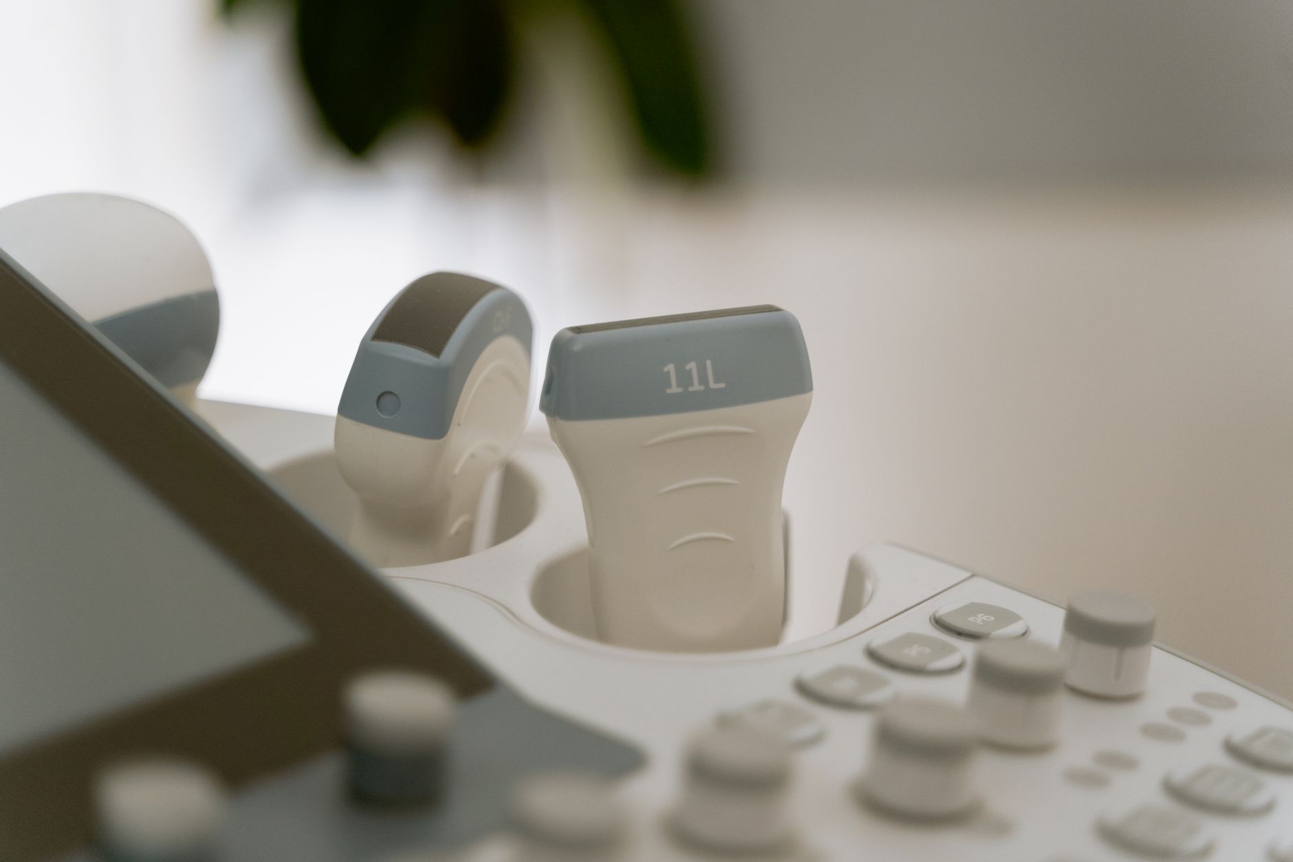
Discover what a sonogram at 7 weeks pregnant reveals, including fetal development, heartbeat detection, and what to expect during your first ultrasound scan.
By Shubhra Mishra — a mom of two who turned her own confusion during pregnancy into BumpBites, a global mission to make food choices clear, safe, and stress-free for every expecting mother. 💛
Check whether any food is safe during pregnancy with the BumpBites Food Safety Checker.
Quick take: A sonogram at 7 weeks pregnant can usually show the gestational sac, yolk sac, tiny embryo and its first heartbeat. The scan is brief—often 10–15 minutes—and is considered safe for both you and your baby. If a heartbeat isn’t seen, your provider will guide the next steps.
It’s 2 a.m., you’re curled up in bed with a warm mug of tea, and the buzzing of a notification pulls you from a half‑finished episode. “What will I see on my 7‑week ultrasound?” you wonder, eyes wide with a mix of excitement and nerves. You’re not alone—most first‑time parents feel the same flutter of anticipation the moment their doctor schedules that early scan.
At seven weeks, the tiny embryo is just beginning to grow, and the ultrasound can give you a visual snapshot of that miracle in motion. In this guide we’ll walk through exactly what a sonogram at 7 weeks pregnant reveals, how the procedure works, what’s considered normal, and what to do if the images look different than expected. We’ll also cover safety, preparation, and the broader schedule of first‑trimester scans so you feel confident every step of the way.
What does a 7‑week pregnancy ultrasound show?
At seven weeks gestation, the ultrasound typically displays four key structures:
Gestational sac – a thin‑walled fluid‑filled space that anchors the pregnancy in the uterine lining.
Yolk sac – a small, round structure that supplies nutrients to the embryo before the placenta forms.
Embryo – the tiny human body, often described as a “bean‑shaped” mass.
Heartbeat – a flickering red dot that can be detected with a Doppler overlay, usually beating between 90–110 beats per minute.
Seeing all four confirms that the pregnancy is progressing as expected. The gestational sac appears first, usually around 4–5 weeks, and by the seventh week the yolk sac and embryo are usually visible. The heartbeat is the most reassuring sign, and it often becomes detectable at this point, though a few days may be needed for the heart to develop enough to be seen.
Can you see a heartbeat at 7 weeks on a sonogram?
Y
es—most clinicians can spot a heartbeat at seven weeks, but the exact timing can vary. The fetal heart begins to beat as early as 5 weeks, yet the ultrasound’s resolution often requires the embryo to be at least 4–5 mm in crown‑rump length (CRL) for the heartbeat to be captured. When visible, the heart rate typically ranges from 90 to 110 bpm.
If the heartbeat isn’t visible on the first scan, doctors usually repeat the exam in 5–7 days. A missed heartbeat at this stage does not automatically mean a problem; the embryo may simply be a few days younger than the estimated gestational age.
What is the size of the embryo at a 7‑week ultrasound?
By the seventh week, the embryo’s crown‑rump length (CRL) measures roughly 5–6 mm. This tiny length is measured from the top of the head (crown) to the bottom of the torso (rump), and it serves as the primary metric for dating the pregnancy. The yolk sac typically measures about 3 mm, while the gestational sac can be 15–20 mm in diameter.
These dimensions can differ slightly based on the exact day of the week and individual growth patterns, but they fall within the ranges endorsed by the American College of Obstetricians and Gynecologists (ACOG) and the UK's Royal College of Obstetricians and Gynaecologists (RCOG).
How long does a 7‑week ultrasound take?
Most 7‑week scans last between 10 and 15 minutes of actual imaging time. Including check‑in, preparation, and the brief post‑scan discussion, the whole appointment usually runs 20–30 minutes. The brevity is one reason many providers schedule these early scans alongside a routine prenatal visit.
Because the embryo is still very small, the sonographer often uses a transvaginal probe, which provides clearer images and can be completed quickly. If an abdominal probe is used, the scan may take a few minutes longer to capture adequate detail.
What are the normal measurements for a 7‑week pregnancy scan?
Standard measurements at seven weeks (± 3 days) are:
Structure
Typical Size
Reference
Embryo (CRL)
5–6 mm
ACOG 2023 Guideline
Yolk sac
≈ 3 mm
RCOG 2022 Recommendations
Gestational sac
15–20 mm diameter
WHO 2021 Ultrasound Standards
Fetal heart rate
90–110 bpm
Mayo Clinic 2024 Review
Measurements falling within these ranges are considered normal. Slight variations are common and usually not a cause for concern, especially if the heart is visible and the embryo appears healthy.
Is it safe to have a sonogram at 7 weeks pregnant?
Yes. Both the American College of Radiology (ACR) and the FDA classify diagnostic obstetric ultrasound as a non‑ionizing, low‑risk procedure when used appropriately. The energy emitted is far below thresholds that could cause tissue heating or cavitation.
Guidelines recommend limiting exposure time—typically under 30 minutes per session—to the minimum needed for diagnostic purposes. No known long‑term adverse effects have been linked to routine early‑pregnancy ultrasounds when performed by qualified professionals.
What to expect during a 7‑week prenatal ultrasound?
When you arrive for your 7‑week scan, the sonographer will first confirm your identity and ask about any recent symptoms. You’ll be asked to wear a hospital gown or a loose top that can be lifted if a transvaginal approach is needed.
During a transvaginal ultrasound, a thin, lubricated probe is gently inserted into the vagina. This method yields clearer images of the tiny embryo and is usually quick and well‑tolerated. If your provider prefers an abdominal scan, you’ll lie on an exam table while a gel is applied to your lower abdomen, and the probe glides over the skin.
Throughout the exam, the sonographer may ask you to hold your breath briefly to improve image clarity. Once the images are captured, they’ll show you the screen—many clinics let you view the live picture, which can be a powerful, emotional moment.
Transvaginal probes provide the clearest view of a 7‑week embryo.
When will the gestational sac appear on a 7‑week scan?
The gestational sac typically becomes visible on ultrasound around 4–5 weeks gestation. By the time you reach seven weeks, the sac is well‑defined and measures roughly 15–20 mm across. Its presence confirms that the pregnancy is implanted within the uterine lining and helps clinicians verify the gestational age.
If the sac isn’t seen at seven weeks, the provider may repeat the scan in a few days or recommend a blood test for human chorionic gonadotropin (hCG) levels to ensure the pregnancy is progressing.
Difference between transvaginal and abdominal ultrasound at 7 weeks
Both approaches are safe, but they differ in image quality and comfort:
Aspect
Transvaginal
Abdominal
Image clarity
High resolution, especially for tiny structures
Lower resolution at early gestation
Discomfort
Brief insertion, usually well‑tolerated
No insertion; may feel cold
Typical use
Preferred ≤ 8 weeks or when early dating is needed
Used later in pregnancy or when transvaginal is contraindicated
Duration
5‑10 minutes of imaging
10‑15 minutes of imaging
Because the embryo is still very small at seven weeks, most clinicians opt for the transvaginal route to ensure the yolk sac and heartbeat are clearly visualized.
Early pregnancy ultrasound images at 7 weeks
Images from a 7‑week scan often look like a tiny bean‑shaped embryo nestled inside a fluid‑filled sac, with a bright red dot indicating the heartbeat. The yolk sac appears as a small, circular structure attached to the embryo’s side. These pictures can be strikingly beautiful, and many parents choose to keep a printed copy as a keepsake.
If you’re curious about what the images might look like, most reputable health websites (e.g., NHS, ACOG) provide sample scans. Remember, each pregnancy is unique, and your scan may look slightly different but still be perfectly normal.
What if no heartbeat is seen at a 7‑week ultrasound?
Finding no heartbeat at seven weeks can be unsettling, but it isn’t always a definitive sign of trouble. Possible reasons include:
The embryo may be a few days younger than estimated, meaning the heart hasn’t yet grown enough to be seen.
Technical factors such as suboptimal positioning or a less‑sensitive probe.
Rarely, a missed miscarriage (also called a “blighted ovum”).
In most cases, the provider will schedule a repeat scan in 5–7 days. If the heartbeat remains absent and the gestational sac does not grow as expected, further evaluation—often including a quantitative hCG test—will be recommended.
How accurate is dating at a 7‑week ultrasound?
Dating based on crown‑rump length at seven weeks is considered highly accurate, typically within ± 5 days of the actual conception date. This level of precision surpasses dating methods that rely solely on the last menstrual period (LMP), especially for women with irregular cycles.
Accurate dating is crucial for scheduling later prenatal screenings, such as the nuchal translucency scan at 11–14 weeks, and for estimating the due date. If the ultrasound measurement differs significantly from the LMP estimate, clinicians will usually adopt the ultrasound dating as the primary reference.
Can a 7‑week ultrasound detect twins?
Yes—if there are two distinct gestational sacs, each with its own yolk sac and embryo, the sonographer can identify a twin pregnancy at seven weeks. However, if the twins share a single sac (monochorionic twins), they may appear as a single sac with two embryos, which can be more challenging to differentiate early on.
Detecting multiples early helps providers plan more frequent monitoring and discuss specific care considerations, such as the increased risk of preterm labor.
What foods to avoid before a 7‑week sonogram?
There are no strict dietary restrictions before a diagnostic ultrasound. However, a light meal can help you feel comfortable during the short exam. Some patients choose to avoid heavy, greasy foods that might cause bloating or discomfort, especially if an abdominal scan is planned.
If you’re scheduled for a transvaginal scan, emptying your bladder beforehand is usually recommended, so you won’t need to drink a large amount of water beforehand as you would for an abdominal exam.
How often should you have ultrasounds in the first trimester?
Most clinicians recommend one early ultrasound—often between 6 and 9 weeks—to confirm the pregnancy, establish dating, and check for multiples. Additional scans may be ordered if:
There are symptoms such as bleeding or severe cramping.
The initial scan couldn’t locate a heartbeat.
There is a history of ectopic pregnancy or other complications.
Beyond the early scan, the next routine ultrasound typically occurs at the anatomy scan around 18‑20 weeks, unless your provider advises otherwise based on your health history.
Enjoy a light snack before your scan—berries provide gentle energy without causing discomfort.
From our medical team: A 7‑week sonogram is a reassuring milestone. Seeing the gestational sac, yolk sac, embryo and heartbeat confirms that your body is nurturing a new life. If any element isn’t visible, remember that timing, positioning and technical factors can influence the images. Your provider will guide you through any needed follow‑up, and most pregnancies progress normally after this early scan.
Myth vs. fact
Myth: You can’t see a baby’s heartbeat until the second trimester. Fact: A heartbeat is often visible on a sonogram as early as seven weeks, though a few days may be needed for the heart to grow enough to be detected.
Myth: Ultrasound radiation can harm the developing embryo. Fact: Diagnostic ultrasounds use non‑ionizing sound waves, and extensive research by the FDA and ACR confirms they are safe when used appropriately.
Myth: You must fast for an early pregnancy scan. Fact: No special fasting is required; a light, comfortable meal is fine, and an empty bladder is often recommended for transvaginal exams.
Key takeaways
A sonogram at 7 weeks pregnant usually shows the gestational sac, yolk sac, embryo and its first heartbeat.
Typical embryo size (crown‑rump length) is 5–6 mm; yolk sac about 3 mm; gestational sac 15–20 mm.
Transvaginal ultrasound provides the clearest images at this stage and is the most common approach.
Early ultrasounds are safe; limit exposure to the shortest time needed for diagnosis.
If no heartbeat is seen, a repeat scan in a week is standard practice.
Dating based on the 7‑week scan is highly accurate and helps schedule later prenatal care.
Frequently asked questions
Can you see the baby’s heartbeat at 7 weeks?
Yes—most clinicians can detect a heartbeat at seven weeks, typically ranging from 90 to 110 bpm. If it isn’t visible on the first scan, a repeat exam in a few days is usually recommended.
How big is the embryo at 7 weeks?
At seven weeks, the embryo’s crown‑rump length measures about 5–6 mm, roughly the size of a sesame seed. The yolk sac is around 3 mm, and the gestational sac measures 15–20 mm in diameter.
What is the gestational sac and when does it appear?
The gestational sac is a fluid‑filled space that anchors the pregnancy in the uterine lining. It generally becomes visible on ultrasound around 4–5 weeks and is well‑defined by the seventh week.
Is a transvaginal ultrasound required at 7 weeks?
While not mandatory, a transvaginal probe is preferred at seven weeks because it offers superior resolution for tiny structures like the yolk sac and early heartbeat.
How accurate is the due date after a 7‑week scan?
Dating based on crown‑rump length at seven weeks is highly accurate, usually within ± 5 days of the true conception date, making it more reliable than dating from the last menstrual period alone.
What are the risks of having an ultrasound at 7 weeks?
Diagnostic ultrasounds use low‑energy sound waves and have no known long‑term risks when performed by qualified professionals. The main precaution is to keep the scan duration as short as reasonably possible.
When to call your doctor
If you experience any of the following after your scan, contact your provider promptly: severe abdominal pain, heavy bleeding, fever, loss of pregnancy symptoms (e.g., nausea) that were previously present, or if you were told the heartbeat was absent and you haven’t had a follow‑up appointment.
These guidelines are for informational purposes only and do not replace personalized medical advice. Always discuss your specific situation with your obstetrician or midwife.
References
American College of Obstetricians and Gynecologists (ACOG). “Ultrasound Guidelines for Obstetric Care,” 2023.
Royal College of Obstetricians and Gynaecologists (RCOG). “Early Pregnancy Ultrasound Recommendations,” 2022.
World Health Organization (WHO). “Standardized Ultrasound Protocols for First Trimester,” 2021.
Mayo Clinic. “Fetal Heart Rate: What’s Normal?” 2024.
Food and Drug Administration (FDA). “Diagnostic Ultrasound Safety,” 2023.
National Institute for Health and Care Excellence (NICE). “Ultrasound in Pregnancy,” 2022.
Centers for Disease Control and Prevention (CDC). “Prenatal Care Guidelines,” 2023.
National Health Service (NHS). “Early Pregnancy Scans,” 2024.
Not sure about the label on 7 Week Pregnancy Ultrasound What You Ll See On The Scan products?
Snap the ingredients list and SafeFilter checks every ingredient for your stage — only 3 free scans this month, then you're locked until reset. Unlimited from $7/mo or lock $50/yr through Aug 31 (6 days left).
When Shubhra Mishra was expecting her first child in 2016, she was overwhelmed by conflicting food advice — one site said yes, another said never. By the time her second baby arrived in 2019, she realized millions of mothers face the same confusion.
That sparked a five-year journey through clinical nutrition papers, cultural diets, and expert conversations — all leading to BumpBites: a calm, compassionate space where science meets everyday motherhood.
Her long-term vision is to build a global community ensuring safe, supported, and free deliveriesfor every mother — because no woman should face pregnancy alone or uninformed. 🌿
🌍 Stand with mothers, shape safer guidance
Join a small circle of experts who review BumpBites articles so expecting parents everywhere can decide with confidence.