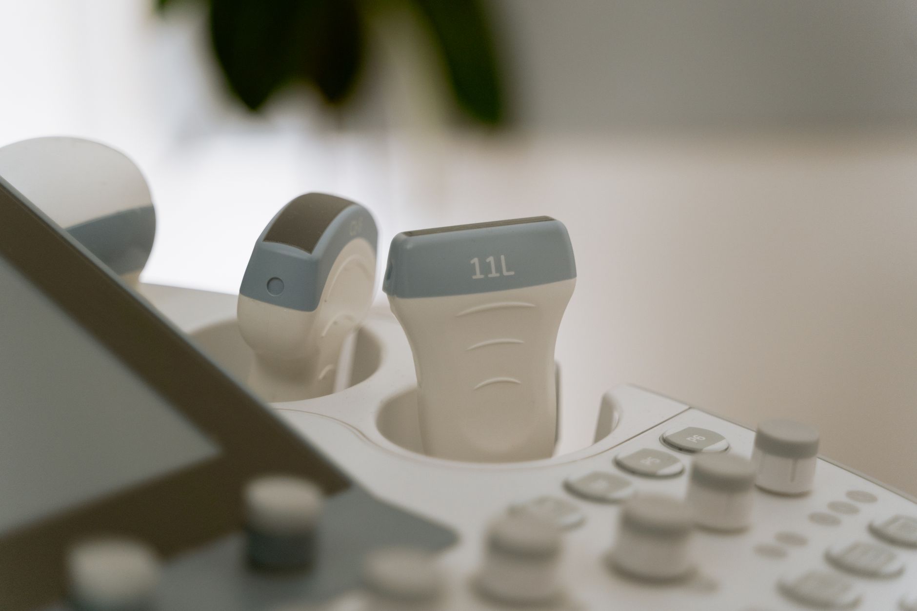
At 6 weeks pregnant, your sonogram pics will likely show a gestational sac, yolk sac, and fetal pole. You might even see a tiny heartbeat! Learn what to expect during your early ultrasound.
By Shubhra Mishra — a mom of two who turned her own confusion during pregnancy into BumpBites, a global mission to make food choices clear, safe, and stress-free for every expecting mother. 💛
Check whether any food is safe during pregnancy with the BumpBites Food Safety Checker.
Quick take: A 6‑week pregnancy ultrasound is safe, brief, and usually shows a tiny gestational sac with a faint heartbeat. It confirms the pregnancy’s location, estimates dating, and helps rule out early complications. If a heartbeat isn’t visible, don’t panic—your provider will likely repeat the scan in a few days.
It’s 2 a.m., you’ve just felt a faint flutter and your partner’s text says, “Did we just hear a baby?” Your mind races to Google: “6 weeks pregnant sonogram pics.” The answer isn’t a mystery you need to solve alone. A 6‑week ultrasound is the first visual glimpse many parents get of their tiny new family member, and it answers a handful of crucial questions while reassuring nervous hearts.
In this article we’ll walk you through exactly what happens at a 6‑week scan, what you’ll likely see on the screen, how accurate the images are, and how to prepare. We’ll also compare it to later scans, discuss costs, and cover safety. By the end you’ll know what to expect, what to ask your provider, and when to seek help.
What to expect at a 6‑week pregnancy ultrasound
At six weeks gestation the embryo is roughly the size of a grain of rice—about 2–4 mm. The ultrasound technician (often called a sonographer) will place a cold gel on your lower abdomen and glide a transducer over the area. The machine sends high‑frequency sound waves that bounce off tissues and create a real‑time image on a monitor. The room is usually dim, quiet, and designed to be a calm space, allowing you to focus on the screen if you wish.
Typical findings include:
Gestational sac: A fluid‑filled sphere that marks the implantation site.
Yolk sac: A tiny circular structure inside the sac that supplies nutrients.
Fetal pole: The earliest visible part of the embryo, appearing as a tiny “dot.”
Heartbeat: A faint flicker, usually detectable around 5.5–6 weeks, with a rate of 90–110 beats per minute.
The scan lasts 5–10 minutes. You’ll hear a soft “whoosh” as the transducer moves, and the screen may show a gray‑scale image that looks like a tiny dot inside a dark circle. If the sonographer spots a heartbeat, they’ll note the rate and often capture a short video clip for your records. This initial contact is often the first time the pregnancy truly feels "real" for many parents, transforming it from a concept to a visible, beating presence.
Most patients leave the office feeling a mix of relief and wonder. One mother we spoke with described the moment the heartbeat appeared: “I heard a tiny ‘thump’ on the speaker and felt my own heart skip. It was the smallest thing that felt so massive.” Beyond the visual, clinics often use the 6‑week visit to review your medical history, confirm your last menstrual period (LMP), and discuss any early symptoms you may be experiencing. This early contact helps set the tone for prenatal care and gives you a chance to voice concerns about nutrition, medications, or lifestyle changes.
What a typical 6‑week scan looks like: a small gestational sac with a faint heartbeat.
6 weeks pregnant sonogram pics and videos
When you search for “6 weeks pregnant sonogram pics,” you’ll encounter a range of images—from textbook diagrams to actual patient videos. Most reputable sites (e.g., Mayo Clinic, NHS) show a gray‑scale picture of the gestational sac with the yolk sac and fetal pole. The heartbeat video often appears as a tiny flickering dot that can be difficult to see without a magnified view, sometimes described as a small, rapid shimmer within the fetal pole. It’s a subtle but profound visual for expecting parents.
Because the embryo is so small, the image quality can vary based on the transducer’s frequency, the mother’s body habitus, and the technician’s skill. High‑frequency (≥ 7 MHz) probes give clearer detail but penetrate less deeply, which is why early scans are performed transabdominally (outside the body) rather than transvaginally unless needed. The sonographer will often point out the key structures on the screen, helping you understand what you're seeing in the often-abstract grayscale image.
If you’re hoping to capture a keepsake, ask the clinic if they can provide a short video clip of the heartbeat. Many providers offer a digital copy for a small fee, and it can be a treasured memento for future family albums. Sharing these first glimpses with close family can be a joyous experience, but remember that the image is very early and abstract, so manage expectations for those who might be hoping to see a fully formed baby.
When reviewing images online, keep in mind that lighting, screen resolution, and image compression can make the heartbeat appear fainter than it was in the clinic. Always compare the video you receive from your provider rather than relying on random internet clips for reassurance, and remember that every scan is unique.
Is a 6‑week ultrasound internal or external?
The term “internal” typically refers to a transvaginal ultrasound, where the probe is inserted into the vagina. At six weeks, most clinicians start with a transabdominal (external) scan because it’s less invasive and usually sufficient to see the gestational sac and heartbeat. This involves the sonographer moving the transducer across your lower abdomen, often with a little pressure to get a clear view.
However, if the baby’s location is unclear, if there’s a history of ectopic pregnancy, or if the ultrasound technician cannot locate a heartbeat, a transvaginal scan may be recommended. Transvaginal ultrasounds provide higher resolution because the probe is closer to the uterus, making it easier to detect very early structures like the fetal pole and heartbeat. While it might sound daunting, the probe is thin, covered with a condom-like sheath, and lubricated for comfort; many women find it less uncomfortable than expected.
Both methods are safe. The International Society of Ultrasound in Obstetrics and Gynecology (ISUOG) states that ultrasound exposure, whether transabdominal or transvaginal, does not pose known risks when used appropriately. ACOG’s Practice Bulletin 225 (2022) recommends starting with transabdominal imaging unless specific risk factors dictate a transvaginal approach. Your comfort and privacy are always prioritized during either procedure.
Insurance plans in the U.S. typically cover the initial transabdominal scan; a transvaginal scan may require prior authorization if it’s not deemed medically necessary, though it's often covered if medically indicated. In the UK, NHS protocols follow NICE guideline NG162, which advises a transabdominal scan first, reserving transvaginal for cases where the pregnancy cannot be visualized or a clearer image is needed for diagnosis.
Can you see the baby's heartbeat at 6 weeks ultrasound?
Yes—most 6‑week scans capture a faint heartbeat. The fetal heart begins to beat around day 22 after conception, but the sound is usually audible on the monitor by 5.5–6 weeks gestation. The typical rate at this stage is 90–110 bpm, slower than the 120–160 bpm seen later in the first trimester. While it's a "flicker" on screen, many clinics will also let you hear the distinct, rapid "thump-thump" sound, which can be an incredibly moving moment.
If a heartbeat isn’t visible, it doesn’t automatically mean a problem. Factors that can obscure the beat include:
Incorrect dating (the pregnancy may be earlier than thought).
Maternal body habitus (higher BMI can reduce image clarity).
Technical limitations of the machine or the angle of the embryo.
In such cases, providers often schedule a repeat scan in 5–7 days to allow the embryo more time to develop and grow, increasing the likelihood of detecting the heartbeat. This waiting period can be anxious, but it's a standard and safe approach. Doppler technology, which adds color flow overlay, can sometimes highlight a faint beat that grayscale imaging misses, but it is used selectively to avoid unnecessary exposure, especially in very early pregnancy.
6‑week ultrasound: what can you see?
Beyond the gestational sac, yolk sac, fetal pole, and heartbeat, a 6‑week scan can provide additional clues:
Uterine location: Confirms the pregnancy is intrauterine, ruling out ectopic pregnancy—a serious condition where the embryo implants outside the uterus.
Number of sacs: Detects multiple gestational sacs, suggesting twins or higher-order multiples, although a definitive count might be clearer at a slightly later scan.
Early signs of viability: A clear yolk sac and a steady heart rate are reassuring markers of a healthy, developing pregnancy.
At this stage you won’t see facial features, limbs, or a recognizable shape. The embryo appears as a tiny “dot” within the sac, often resembling a small grain of rice. It's important to set realistic expectations; you won't be seeing a "baby" in the traditional sense, but rather the earliest, fundamental building blocks of one. Later ultrasounds (around 12 weeks) will reveal a more defined silhouette and the beginnings of recognizable body parts.
Some clinicians also assess the thickness of the uterine lining (endometrium) and note any fluid collections that could hint at early complications. While these observations are subtle, they help form a baseline for later comparison and provide your healthcare team with a comprehensive early picture of your pregnancy.
How accurate is a 6‑week pregnancy ultrasound?
When performed by a trained sonographer using a high‑quality machine, a 6‑week scan is highly accurate for confirming intrauterine pregnancy and estimating gestational age within ± 3–5 days. This early dating is crucial because it helps establish your estimated due date, which guides all subsequent prenatal care, screenings, and interventions. The American College of Obstetricians and Gynecologists (ACOG) notes that dating accuracy improves with each subsequent scan, but the early scan is reliable enough to guide first‑trimester care and set a precise timeline for your pregnancy journey.
Accuracy can be affected by:
Incorrect last menstrual period (LMP) dating, making the ultrasound dating even more valuable.
Maternal obesity, which can sometimes reduce the clarity of the image.
Suboptimal equipment or the skill level of the sonographer.
Even with these variables, a correctly performed scan will reliably detect a gestational sac, yolk sac, fetal pole, and heartbeat, providing essential reassurance for most patients. Studies published in the *Journal of Ultrasound in Medicine* (2021) found that 94 % of scans at 6 weeks correctly identified the fetal pole when the LMP was within a 7‑day window, underscoring its reliability. This early confirmation helps alleviate much of the initial uncertainty and anxiety that often accompanies early pregnancy.
What if no heartbeat is seen at a 6‑week ultrasound?
Seeing no heartbeat can feel incredibly alarming and frightening, but it’s not a definitive sign of miscarriage. It’s a situation that requires careful follow-up and further assessment. Common reasons include:
Dating error: The pregnancy may be younger than 6 weeks. If your ovulation or conception happened later than you thought, the embryo simply hasn't developed enough yet. A repeat scan in a week often resolves the issue.
Technical factors: High maternal BMI, an unfavorable fetal position, or even a slightly retroverted uterus can sometimes hide the beat, making it difficult to visualize at this very early stage.
Early loss: In some cases, unfortunately, the embryo may not be viable, and a follow‑up scan will confirm a miscarriage.
What to do:
Stay calm and schedule a repeat ultrasound in 5–7 days. This waiting period, though difficult, is essential for accurate diagnosis.
Ask your provider about possible blood tests (e.g., quantitative hCG) to assess viability, as rising hCG levels can be reassuring.
Monitor for symptoms like cramping or bleeding, and report any sudden changes to your medical team immediately.
Most clinicians view a single absent heartbeat at six weeks as a “re‑scan” situation rather than an immediate cause for concern. Emotional support is also key; many practices offer counseling or a follow‑up phone call to address anxiety and help you navigate this uncertain period. Remember, you're not alone in this experience.
6‑week ultrasound risks and complications
Ultrasound is considered a non‑invasive, low‑risk imaging modality. The U.S. Food and Drug Administration (FDA) and the UK National Institute for Health and Care Excellence (NICE) classify diagnostic obstetric ultrasound as safe when used appropriately. Reported complications are extremely rare and include:
Minor skin irritation from the gel, which is typically hypoallergenic.
Transient discomfort from the transducer pressure, especially if a clearer view is needed.
In the case of transvaginal scans, a brief moment of mild discomfort, similar to a pelvic exam.
There is no evidence that the low‑intensity sound waves used in diagnostic ultrasound cause harm to the embryo. Decades of research and widespread use support its safety profile when performed by qualified professionals. However, ACOG advises that ultrasound should be performed only when medically indicated, avoiding “keepsake” scans solely for non‑clinical reasons, as these can lead to unnecessary exposure or false reassurance without medical oversight.
Guidelines recommend limiting the total exposure time to the shortest duration necessary for a diagnostic answer. The ISUOG consensus (2021) emphasizes that even repeated scans within the first trimester have not shown adverse fetal outcomes when protocols are followed. Your provider will always weigh the benefits of the scan against any theoretical risks, ensuring your and your baby's safety.
Why an early 6‑week ultrasound is performed (beyond routine)
While some practices include an early ultrasound as a routine part of prenatal care, a 6-week scan is often specifically recommended for particular medical reasons. These indications help your provider gather crucial information early on, ensuring a safer and more informed pregnancy journey.
Common reasons for an early ultrasound include:
Uncertainty about dating: If you're unsure of your last menstrual period (LMP) or have irregular cycles, an early scan provides the most accurate estimated due date based on the embryo's size.
Vaginal bleeding or spotting: Any bleeding in early pregnancy can be alarming. An ultrasound can help determine the cause, rule out serious issues like ectopic pregnancy, or confirm the presence of a healthy, intrauterine pregnancy.
Abdominal pain: Similar to bleeding, pain can be a symptom of various conditions, including ectopic pregnancy, ovarian cysts, or early miscarriage. An ultrasound helps pinpoint the source.
History of complications: If you've had a previous ectopic pregnancy, recurrent miscarriages, or fertility treatments like IVF, an early scan is vital to confirm proper implantation and viability.
Concerns about multiples: If you've undergone fertility treatments or have a family history of twins, an early scan can detect multiple gestational sacs, indicating the possibility of a multiple pregnancy.
In these situations, the 6-week ultrasound is not just a glimpse of your baby; it's a critical diagnostic tool that guides your care and provides essential reassurance or identifies potential issues that need immediate attention.
Understanding your 6‑week ultrasound measurements
At your 6-week ultrasound, the sonographer will take a few key measurements that help your doctor assess the pregnancy's progression and confirm dating. These measurements, though tiny, are crucial indicators of early development.
Here’s what they typically measure and what the numbers mean:
Measurement
Description
Typical Range at 6 Weeks
Significance
Gestational Sac Diameter (GSD)
The size of the fluid-filled sac surrounding the embryo.
10-18 mm
Indicates the presence of an intrauterine pregnancy and helps with early dating.
Crown-Rump Length (CRL)
The length of the embryo from the top of its head (crown) to its bottom (rump).
2-4 mm
The most accurate measurement for dating in early pregnancy, typically within 3-5 days.
Fetal Heart Rate (FHR)
The number of times the embryo's heart beats per minute.
90-110 bpm
A key indicator of viability and healthy development at this stage.
These measurements provide your healthcare team with objective data, complementing the visual assessment of the gestational sac, yolk sac, and fetal pole. While these numbers are important, remember that early development can vary slightly, and your provider will interpret them in the context of your overall health and pregnancy history. Don't hesitate to ask your sonographer or doctor to explain what each measurement signifies for your pregnancy.
What happens after your 6‑week ultrasound? Next steps in prenatal care
After the excitement and perhaps anxiety of your 6-week ultrasound, you might be wondering what comes next. This early scan is just the first step in a comprehensive prenatal care plan designed to support a healthy pregnancy. Your provider will discuss the findings with you, often immediately after the scan or at a follow-up appointment.
Here's what you can generally expect in the days and weeks following your 6-week ultrasound:
Review of results: Your doctor or midwife will go over the ultrasound report, confirming the pregnancy's location, estimated due date, and viability markers like the heartbeat. This is your opportunity to ask any questions that came up during or after the scan.
First prenatal appointment: If you haven't already had it, your provider will schedule your first comprehensive prenatal visit. This appointment typically involves a physical exam, blood tests (to check blood type, iron levels, infection screening), urine tests, and a detailed discussion about your medical history, lifestyle, and any concerns you have.
Prenatal vitamins and nutrition: You'll likely receive or reiterate guidance on continuing or starting prenatal vitamins, especially those containing folic acid, and advice on healthy eating during pregnancy.
Lifestyle adjustments: Discussions about safe exercise, avoiding certain foods, and managing common early pregnancy symptoms like nausea or fatigue will be part of your ongoing care.
Planning for future scans: Your provider will outline the schedule for subsequent ultrasounds, such as the 12-week nuchal translucency scan (often combined with blood tests for genetic screening) or the detailed anatomy scan around 18-22 weeks.
Emotional support: Pregnancy is a time of immense change. Your care team can also connect you with resources for emotional support, whether it's for managing anxiety, navigating relationship changes, or preparing for parenthood.
This period is about establishing a strong foundation for your pregnancy, ensuring you have all the information and support you need as your baby continues to grow.
Capturing the first heartbeat: a gentle probe and a quiet moment of connection.
How body type and uterine position affect the 6‑week scan
Maternal body habitus plays a significant role in image quality. Higher body mass index (BMI) can increase the distance between the transducer and the uterus, reducing resolution and sometimes obscuring the fetal pole or heartbeat. This doesn't mean there's a problem with the pregnancy; it simply means the sonographer might need to make adjustments to get a clear view. In such cases, sonographers may adjust the probe frequency, use a slightly higher‑gain setting on the machine, or recommend a transvaginal scan for clearer visualization, as it bypasses the abdominal wall.
Uterine position—whether the uterus is tilted forward (anteverted) or backward (retroverted)—also influences how easily the embryo is seen. An anteverted uterus, which tilts towards the abdomen, often provides a clearer transabdominal view. Conversely, a retroverted uterus can place the gestational sac farther from the abdominal wall, making a transabdominal view more challenging. ACOG’s guidelines suggest that when the uterus is difficult to image transabdominally, a transvaginal approach is appropriate and safe, providing a clearer picture without additional risk. Your sonographer is skilled in these adjustments and will always aim for the best possible image while ensuring your comfort.
Emotional experience: what to expect after seeing the heartbeat
Seeing the tiny flicker of a heartbeat often triggers a wave of emotions: joy, relief, awe, and sometimes unexpected anxiety about the next steps. It’s normal to feel a mix of excitement and nervousness. Couples frequently describe the moment as “the first real proof” that a baby is growing, which can also bring up worries about health, lifestyle changes, and future appointments. This profound experience can solidify the reality of pregnancy in a way that a positive test never could.
Take a minute after the scan to process those feelings. Many clinics have a short debrief period where the sonographer or provider will explain what was seen, answer immediate questions, and suggest next‑step milestones. If you feel overwhelmed, consider bringing a supportive partner, friend, or family member, and schedule a brief follow‑up call if you have lingering concerns. It’s also perfectly normal for partners to experience a similar range of emotions, moving from abstract anticipation to concrete connection. Allow yourselves to feel and share these moments together.
From our medical team: A 6‑week scan is a reassuring first checkpoint. It confirms the pregnancy is where it should be, gives you an early glimpse of your baby’s heartbeat, and helps your provider plan prenatal care. If anything looks unclear, a repeat scan is standard practice—not a cause for alarm. Trust your medical team to guide you through this exciting and sometimes uncertain time.
Myth vs. fact
Myth: You must see a clear baby face at six weeks.
Fact: Facial features develop later; at six weeks the embryo looks like a tiny dot, not a recognizable baby. Realistic expectations are key for this early scan.
Myth: All early ultrasounds are risky and can harm the baby.
Fact: Diagnostic ultrasound uses low‑intensity sound waves and is considered safe when performed by a qualified professional for medical reasons. There's no evidence of harm.
Myth: If you don’t see a heartbeat, the pregnancy is lost.
Fact: The heartbeat may be too early to detect due to dating errors or technical factors; a repeat scan in a few days often resolves the question, offering clarity and reassurance.
Myth: An early ultrasound can determine the baby's gender.
Fact: At six weeks, the external genitalia are not yet developed enough to be visible on an ultrasound. Gender determination is typically possible much later, around 18-22 weeks.
Key takeaways
A 6‑week ultrasound safely confirms an intrauterine pregnancy and usually shows a gestational sac, yolk sac, fetal pole, and faint heartbeat.
Preparation is simple: drink enough water, wear comfortable clothing, and bring any questions you have.
If a heartbeat isn’t visible, a repeat scan in 5–7 days is the typical next step, not an immediate cause for alarm.
Risks are minimal; the only real discomfort may be brief pressure from the transducer or mild discomfort during a transvaginal scan.
Costs vary by region and insurance status; private US clinics charge $150‑$300, while NHS scans are free.
Later scans (8 weeks and beyond) reveal more anatomy and provide clearer heart‑rate data.
Body type and uterine position can affect image clarity; your provider will adjust technique as needed to get the best view.
Seeing the heartbeat often brings strong emotions—give yourself time to process and lean on support from your partner and medical team.
Early ultrasounds are often performed for specific medical reasons, such as bleeding, pain, or uncertain dating, providing crucial diagnostic information.
Frequently asked questions
Can you see the baby's face at a 6‑week ultrasound?
No. At six weeks the embryo is too small to show facial features; it appears as a tiny dot within the gestational sac. You'll need to wait for later scans to see more defined features.
How big is the baby at a 6‑week ultrasound?
The embryo measures about 2–4 mm—roughly the size of a grain of rice—and is nestled inside a fluid‑filled sac. This tiny size is why it appears as a small "flicker" on the screen.
What is the purpose of a 6‑week pregnancy ultrasound?
The main goals are to confirm that the pregnancy is inside the uterus, estimate gestational age, detect a heartbeat, and rule out early complications such as ectopic pregnancy or multiple gestations.
Can a 6‑week ultrasound detect twins?
It can identify multiple gestational sacs, which suggests twins or higher‑order multiples, but a definitive diagnosis with individual fetal poles and heartbeats may require a follow‑up scan at 8‑10 weeks for clearer visualization.
Is a 6‑week ultrasound necessary?
While not mandatory for every pregnancy, many clinicians recommend it to confirm dating, ensure viability, and establish a baseline for prenatal care, especially if there are specific concerns or an unclear LMP.
Can I get a 3D ultrasound at six weeks pregnant?
3D imaging at six weeks is limited because the embryo is very small and lacks developed features; the technology becomes more useful after 10‑12 weeks when structures are larger and more defined for detailed visualization.
What if my period is late but I can't find a heartbeat at 6 weeks?
Late periods can mean the gestational age is earlier than expected, making the heartbeat harder to detect. Your provider will likely order a repeat scan in a few days and may check quantitative hCG levels to assess viability, providing clarity over time.
Can a 6‑week ultrasound detect multiple pregnancies?
Early scans can reveal more than one gestational sac, indicating the possibility of twins or higher-order multiples. However, the exact number of embryos and their individual heartbeats may not be definitively clear until a later scan (around 8–10 weeks) when they have grown larger.
Is bleeding or cramping normal before a 6‑week ultrasound?
Light spotting can sometimes be normal, such as implantation bleeding, but heavy bleeding or severe cramping should always be reported to your doctor immediately. While not always a sign of a problem, these symptoms warrant prompt medical evaluation to rule out complications.
What if I don't know my last menstrual period (LMP) for dating?
If you don't know your LMP, the 6-week ultrasound becomes even more valuable. The crown-rump length (CRL) measurement taken at this stage is highly accurate for establishing your estimated due date, often within a few days, providing essential clarity for your prenatal care plan.
When to call your doctor
If you experience any of the following after your scan, contact your provider promptly: heavy vaginal bleeding (more than spotting), severe abdominal pain or cramping, fever over 100.4 °F (38 °C), or a sudden loss of fetal movement (though at six weeks movement is rarely felt). Remember, this article is for information only and does not replace personal medical advice from your healthcare provider.
References
American College of Obstetricians and Gynecologists (ACOG). “Ultrasound in Pregnancy.” Practice Bulletin No. 225, 2022.
National Health Service (NHS). “Early Pregnancy Ultrasound.” Updated 2023.
Mayo Clinic. “First‑Trimester Ultrasound.” Patient Care Information, 2023.
International Society of Ultrasound in Obstetrics and Gynecology (ISUOG). “Safety of Diagnostic Ultrasound.” Consensus Statement, 2021.
U.S. Food and Drug Administration (FDA). “Ultrasound Safety.” Guidance Document, 2022.
World Health Organization (WHO). “Recommendations on Antenatal Care for a Positive Pregnancy Experience.” 2022.
National Institute for Health and Care Excellence (NICE). “Ultrasound Scanning in Pregnancy.” Clinical Guideline NG162, 2021.
Centers for Disease Control and Prevention (CDC). “Prenatal Care.” 2023.
Journal of Ultrasound in Medicine. “Accuracy of Early First‑Trimester Ultrasound for Dating.” 2021.
Not sure about the label on 6 Week Pregnancy Ultrasound What You Ll See On The Scan products?
Snap the ingredients list and SafeFilter checks every ingredient for your stage — only 3 free scans this month, then you're locked until reset. Unlimited from $7/mo or lock $50/yr through Aug 31 (6 days left).
When Shubhra Mishra was expecting her first child in 2016, she was overwhelmed by conflicting food advice — one site said yes, another said never. By the time her second baby arrived in 2019, she realized millions of mothers face the same confusion.
That sparked a five-year journey through clinical nutrition papers, cultural diets, and expert conversations — all leading to BumpBites: a calm, compassionate space where science meets everyday motherhood.
Her long-term vision is to build a global community ensuring safe, supported, and free deliveriesfor every mother — because no woman should face pregnancy alone or uninformed. 🌿
🌍 Stand with mothers, shape safer guidance
Join a small circle of experts who review BumpBites articles so expecting parents everywhere can decide with confidence.