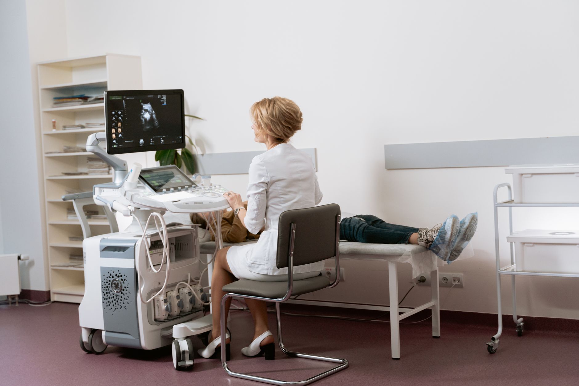
Get answers on 12 Week Pregnancy USG Nuchal Translucency Scan, understand its importance and what to expect during this crucial test
By Shubhra Mishra — a mom of two who turned her own confusion during pregnancy into BumpBites, a global mission to make food choices clear, safe, and stress-free for every expecting mother. 💛
Check whether any food is safe during pregnancy with the BumpBites Food Safety Checker.
Quick take: The 12‑week pregnancy ultrasound, called a nuchal translucency (NT) scan, measures the fluid‑filled space at the back of your baby’s neck to help assess the risk of certain chromosomal conditions. It’s a safe, quick test that’s optional in most settings, but a normal result greatly reduces—but does not eliminate—the chance of Down syndrome or other trisomies.
It’s 2 a.m. and you’ve just felt the tiny flutter of your baby for the first time. A quick Google search later, you’re staring at the phrase “12 week pregnancy USG nuchal translucency scan” and wondering whether that early‑pregnancy ultrasound is something you must have, what it actually looks at, and if a weird result could mean trouble. You’re not alone—many expectant parents feel the same mix of excitement and anxiety.
In this guide we’ll explain exactly what the NT scan is, why it’s done at around 12 weeks, what the numbers mean, how it fits with other screening options, and what to expect on the day of the exam. We’ll also cover costs, preparation tips, the test’s accuracy, and the next steps if the measurement is higher than average. By the end you’ll have a clear picture of the benefits, limits, and choices surrounding the 12‑week NT scan, so you can discuss the results confidently with your provider.
What is the 12 week nuchal translucency scan and why is it done?
The nuchal translucency scan is a specialized first‑trimester ultrasound performed between 11 weeks 0 days and 13 weeks 6 days of gestation. During the scan the sonographer measures the thickness of the “nuchal translucency”—a clear fluid‑filled space at the back of the fetus’s neck. This measurement, when combined with maternal blood‑test results (the combined first‑trimester screen), helps estimate the risk of chromosomal abnormalities such as Down syndrome (trisomy 21), trisomy 18, and trisomy 13.
Why focus on the neck? Research shows that embryos with certain chromosomal conditions often have a larger fluid collection in the nuchal region, reflecting altered fetal development. By catching this early, clinicians can offer further testing (non‑invasive prenatal testing, chorionic villus sampling, or amniocentesis) and give families more time to make informed choices.
In the United States, the American College of Obstetricians and Gynecologists (ACOG) recommends offering the combined screen—including the NT measurement—to all pregnant people, but it is not mandatory. In the United Kingdom, NHS guidelines similarly make it an optional part of the first‑trimester screen, with the decision left to the individual after counseling.
Beyond chromosomal risk, the NT scan also provides a quick anatomical “first look” at the fetus. The sonographer can assess the fetal head shape, facial profile, and early cardiac activity, which can flag structural concerns that might need a follow‑up anatomy scan later in the second trimester.
During a 12‑week NT scan the sonographer captures a clear image of the fetal neck to measure fluid thickness.
What does a nuchal translucency measurement indicate at 12 weeks?
A single NT measurement does not diagnose a condition; it indicates risk. The thicker the translucency, the higher the statistical chance that the fetus carries a chromosomal abnormality. The measurement is expressed in millimeters (mm) and is interpreted alongside two key pieces of information:
Gestational age: The exact age of the pregnancy (based on the first day of the last menstrual period or an early‑dating scan) is crucial because the NT thickness naturally changes as the fetus grows.
Maternal serum markers: Levels of pregnancy‑associated plasma protein‑A (PAPP‑A) and free beta‑human chorionic gonadotropin (β‑hCG) measured from a blood draw at the same visit. When combined, these values generate a risk percentage for each condition.
In practice, a low NT measurement (often ≤ 2.5 mm) paired with normal serum markers yields a low risk—commonly reported as less than 1 in 1,000 for Down syndrome. A higher NT measurement raises the risk, sometimes to 1 in 100 or higher, prompting discussion of additional testing.
Risk calculators also adjust for maternal weight, ethnicity, and smoking status, because these factors can subtly influence serum‑marker levels. The resulting risk percentage is a statistical estimate, not a definitive prediction, and should always be interpreted by a qualified provider.
What is a normal nuchal translucency range at 12 weeks pregnant?
“Normal” is a statistical range rather than an absolute cutoff. Large population studies, such as those summarized in ACOG Practice Bulletin No. 163, show that the median NT thickness at 12 weeks is about 1.4 mm, with most values falling between 1.0 mm and 2.5 mm. Here’s a quick reference:
Gestational Age (weeks)
Typical NT Range (mm)
11 weeks 0 days – 11 weeks 6 days
0.9 – 2.2
12 weeks 0 days – 12 weeks 6 days
1.0 – 2.5
13 weeks 0 days – 13 weeks 6 days
1.2 – 2.9
If the NT measurement falls within these ranges and the serum markers are also in the normal range, the combined risk is usually considered low. However, a measurement slightly above the typical range (e.g., 2.6 mm) does not automatically mean a problem; it simply warrants a closer look.
Many laboratories publish percentile charts (e.g., 95th percentile) that help clinicians decide whether a value is “borderline” or truly elevated. These charts are based on large, diverse cohorts and are updated periodically to reflect evolving data.
What happens if your 12 week NT scan results are high or abnormal?
When the NT measurement is higher than expected, the first step is usually a detailed counseling session with your obstetrician or a maternal‑fetal medicine specialist. They will explain the specific risk percentages for each condition based on your combined results.
Common follow‑up options include:
Non‑invasive prenatal testing (NIPT): A blood test that analyzes fetal DNA fragments circulating in the mother’s bloodstream. NIPT has a detection rate > 99 % for trisomy 21 and a false‑positive rate under 0.1 % (according to the International Society for Prenatal Diagnosis, 2021).
Chorionic villus sampling (CVS): An invasive diagnostic test performed between 10 and 13 weeks that collects placental tissue for chromosomal analysis. It provides a definitive result but carries a small risk of miscarriage (about 0.5 % per ACOG).
Amniocentesis: Usually offered after 15 weeks, this test samples amniotic fluid. It has a slightly lower miscarriage risk than CVS (≈ 0.3 %) but is not typically the first choice when a high NT is found at 12 weeks.
It’s important to remember that a high NT measurement can also be associated with non‑chromosomal outcomes, such as congenital heart defects or genetic syndromes not covered by standard NIPT panels. Therefore, comprehensive counseling should address the full spectrum of possibilities.
Emotional support is a key part of this conversation. Many clinics now offer a brief session with a genetic counselor or a psychologist to help families process anxiety and make decisions that align with their values.
Is the 12 week nuchal translucency scan mandatory or optional?
In most healthcare systems the NT scan is offered as part of the combined first‑trimester screen, but it is not legally required. The decision rests on informed consent:
United States: ACOG states that screening tests, including the NT scan, should be offered to all pregnant people, but accepting or declining is a personal choice.
United Kingdom: NHS guidance lists the combined test as the standard offering; patients can decline after receiving balanced information about benefits and limitations.
Other countries: Many European and Australian guidelines follow a similar opt‑in approach, emphasizing patient autonomy.
Cultural, religious, or personal beliefs sometimes influence the decision to decline. For example, some families prefer to avoid any invasive testing unless absolutely necessary, while others value early information to prepare for potential interventions.
If you choose to skip the NT scan, you can still pursue other screening methods (like NIPT alone) later in pregnancy. However, opting out means you lose the early‑gestation risk estimate that can guide timely decision‑making.
How accurate is the nuchal translucency scan for detecting chromosomal abnormalities?
Accuracy is best understood as a combination of detection rate (sensitivity) and false‑positive rate (specificity). When combined with maternal serum markers, the NT scan detects about 85 % of Down‑syndrome cases and 70–80 % of trisomy 18/13 cases (ACOG 2020). The false‑positive rate for the combined screen is roughly 5 %—meaning that out of 100 pregnancies flagged as high risk, about five will be false alarms.
When used alone (NT measurement without serum markers), sensitivity drops to around 65 % for trisomy 21, underscoring why the combined approach is the standard. Importantly, the test’s predictive value depends heavily on the baseline prevalence of the condition, which varies with maternal age and other risk factors.
Comparing NT to NIPT, the latter has higher sensitivity (> 99 % for trisomy 21) and a lower false‑positive rate (< 0.1 %). However, NIPT is a screening test, not a diagnostic one, and it is typically performed after the 10‑week mark. Many clinicians use the NT scan as an early “first look” and then follow up with NIPT if risk is elevated.
What should you expect during your 12 week nuchal translucency ultrasound appointment?
First, you’ll arrive at the imaging suite and change into a loose, comfortable gown. The sonographer will confirm your gestational age, usually by checking the crown‑rump length (CRL). Then, with a warm gel applied to your abdomen, they’ll place the transducer (the hand‑held probe) gently on your skin.
The scan itself takes about 10–15 minutes. The sonographer looks for the following landmarks:
Clear view of the fetal head in a midsagittal plane.
Visible nasal bone.
The “clear space” at the back of the neck.
Because the NT measurement is so small, the technician may ask you to hold your breath briefly to reduce fetal movement and improve image clarity. After the images are captured, the technician will forward them to a radiologist or maternal‑fetal specialist for measurement and reporting. You’ll typically receive a written result within a few days, along with an interpretation of the combined risk if serum markers were also drawn.
Most patients report feeling only mild pressure from the probe and no discomfort. If you’re nervous, you can bring a partner or support person, ask the sonographer to explain each step, and request a short break if needed. Occasionally, the first scan may not provide a perfect view; in those cases, a repeat scan within a few days is standard practice.
What is the typical cost of a 12 week nuchal translucency scan?
In the United States, private‑pay rates for the NT scan range from $150 to $400, depending on the clinic’s location and whether the measurement is bundled with the combined serum‑marker test. For those with insurance, many plans cover the scan as part of prenatal screening, though co‑pays or deductibles may apply.
In the United Kingdom, the NHS provides the combined first‑trimester screen—including the NT scan—free of charge to residents. Private clinics charge roughly £200‑£300 for the scan alone, with additional fees if you request a detailed report or a second opinion.
Other countries have similar structures: Canada’s provincial health plans generally cover the scan; Australia’s Medicare rebates cover the combined test, while private facilities may charge $200‑$300 AUD. In many Asian markets, the cost can vary widely—from $100 USD in public hospitals to $500 USD in high‑end private centers. Always verify coverage with your insurer or national health service ahead of time to avoid surprise bills.
Additional considerations
NT scan vs. NIPT at 12 weeks
Both tests can be done at 12 weeks, but they serve different roles. The NT scan is an ultrasound‑based risk estimate that also provides a visual of fetal anatomy. NIPT analyzes cell‑free fetal DNA in maternal blood and offers higher specificity for trisomies. Many clinicians recommend the NT scan first because it gives an early anatomical snapshot; if the combined risk is high, NIPT is then ordered for a more precise assessment.
Preparation tips for your NT scan
Come with a full bladder if instructed—some clinics find a partially full bladder improves image quality.
Wear a comfortable, loose‑fitting top that can be easily lifted.
Bring a list of any medications or supplements you’re taking; certain substances can affect serum marker levels.
Plan to stay for at least 30 minutes to allow for the scan and any immediate questions.
False‑positive rate of the NT scan
The combined first‑trimester screen, which includes NT, has a false‑positive rate of about 5 % (ACOG 2020). This means that out of 100 pregnancies flagged as high risk, five will turn out to be normal after diagnostic testing. Understanding this rate helps set realistic expectations and reduces unnecessary anxiety.
Interpreting NT scan results
A result typically includes three parts: the NT measurement in millimeters, the calculated risk for each trisomy, and a recommendation. For example, a report might read “NT = 2.1 mm; combined risk for trisomy 21 = 1 in 850; recommend NIPT.” The risk number is relative; a lower number (e.g., 1 in 10,000) is reassuring, while a higher number (e.g., 1 in 100) signals a need for further evaluation.
NT scan for twins
When a pregnancy involves twins, each fetus is measured separately, and the highest NT value is used for risk calculation. The combined screen still applies, but the overall risk interpretation can be more complex, and some providers may suggest early NIPT for each twin if the NT measurement is elevated.
Risks and benefits of the NT scan
Risks are minimal—ultrasound energy is considered safe, and the scan does not involve radiation. The main benefit is early risk stratification, which can guide decisions about further testing and pregnancy management. The small potential downside is anxiety from false‑positive results, which is why thorough counseling is essential.
Why gestational age matters for the NT scan
Accurate dating is crucial because NT thickness naturally increases as the fetus grows. A scan performed too early (before 11 weeks) may underestimate the measurement, while a scan done after 13 weeks 6 days may overestimate it. That’s why providers verify the dating with a crown‑rump length measurement before taking the NT reading.
Understanding the combined first‑trimester screen
The combined first‑trimester screen merges three pieces of data: the nuchal translucency measurement, the crown‑rump length (to confirm gestational age), and two maternal serum markers (PAPP‑A and free β‑hCG). When entered into a risk algorithm—often provided by the laboratory—the output is a probability that the fetus has trisomy 21, trisomy 18, or trisomy 13. This integrated approach is more powerful than any single component alone, achieving detection rates around 85 % for Down syndrome (ACOG 2020).
Because the calculation accounts for maternal age, weight, ethnicity, and smoking status, the same NT measurement can yield different risk percentages for different patients. For example, a 28‑year‑old non‑smoker with a 2.0 mm NT may have a risk of 1 in 2,500, while a 38‑year‑old smoker with the same NT might have a risk of 1 in 800. Understanding this nuance helps families appreciate why a “high‑risk” result does not always mean a diagnosis, but rather a signal to consider more definitive testing.
How maternal age and other factors influence NT risk assessment
Maternal age remains the strongest single predictor of chromosomal abnormalities. The baseline risk of Down syndrome at age 25 is roughly 1 in 1,250, rising to about 1 in 100 by age 40. When the NT measurement is added, the age‑adjusted risk can shift dramatically. For instance, a 2.5 mm NT at 12 weeks may double the age‑related risk for a 35‑year‑old, but only modestly increase it for a younger mother.
Other modifiers include maternal obesity (higher weight can dilute serum marker concentrations), diabetes (which can raise β‑hCG levels), and certain medications such as fertility drugs. Clinicians often request a repeat serum marker draw if these factors are present, ensuring the combined risk estimate is as accurate as possible.
Options for early non‑invasive prenatal testing (NIPT) at 12 weeks
Non‑invasive prenatal testing can be performed as early as 10 weeks, making it a viable follow‑up after a high‑risk NT screen. NIPT analyzes fragments of fetal DNA that circulate in the mother’s bloodstream, providing a highly sensitive screen for trisomy 21, trisomy 18, trisomy 13, and, in many commercial panels, sex chromosome aneuploidies.
Because NIPT does not involve an invasive procedure, it carries virtually no physical risk to the fetus. However, it remains a screening test; a positive result still requires confirmatory diagnostic testing (CVS or amniocentesis) before any clinical decisions are made. Many insurers now cover NIPT when a combined first‑trimester screen is flagged as high risk, so discuss coverage options with your provider.
Understanding the emotional journey after a high‑risk NT result
Receiving a high‑risk result can trigger a cascade of emotions—shock, fear, and a sense of urgency. It’s normal to feel overwhelmed. Studies show that couples who receive timely counseling and clear information report lower long‑term anxiety (Mayo Clinic, 2022). Most clinics have a genetic counselor on staff who can explain the meaning of risk percentages, outline next‑step options, and provide decision‑making support.
It can also be helpful to write down questions before your next appointment. Examples include: “What is the exact risk for Down syndrome?”, “How does my age affect this risk?”, and “What are the timing and risks of CVS versus amniocentesis?” Having a list ready ensures you get the answers you need without feeling rushed.
Normal (left) versus mildly elevated (right) nuchal translucency measurements on a 12‑week scan.
From our medical team: The 12‑week NT scan is a valuable early‑pregnancy tool, but it should be viewed as part of a broader screening strategy. A normal NT measurement, especially when combined with reassuring serum markers, provides strong reassurance but does not guarantee the absence of all chromosomal conditions. If you receive a high‑risk result, discuss NIPT and diagnostic options with your provider, and remember that many outcomes remain favorable even when the NT is mildly elevated.
Myth vs. fact
Myth: A normal NT scan means there is zero chance of Down syndrome.
Fact: A normal NT measurement significantly lowers the risk, but it does not eliminate it. Even with a low NT, a small residual risk remains, which is why some families still choose NIPT for added reassurance.
Myth: The NT scan is painful and risky for the baby.
Fact: The scan uses harmless sound waves and typically feels like a gentle pressure on the abdomen. There is no known risk to the fetus from a properly performed NT ultrasound.
Myth: You must have the NT scan to be offered any prenatal testing.
Fact: While the NT scan is a common component of the combined first‑trimester screen, other screening options (such as NIPT alone) are available, and the decision is ultimately personal.
Key takeaways
The 12‑week NT scan measures fluid at the back of the fetal neck to assess early risk for trisomies.
Normal NT values are usually ≤ 2.5 mm; higher measurements raise the statistical risk but are not diagnostic.
The scan is optional; you can decline after receiving balanced counseling.
Combined with maternal serum markers, the NT scan detects about 85 % of Down‑syndrome cases with a ~5 % false‑positive rate.
If the result is high, follow‑up options include NIPT, CVS, or amniocentesis—all discussed with your provider.
Preparation is simple—wear comfortable clothing, bring a list of medications, and arrive with a partially full bladder if asked.
Costs vary by country and insurance; many public health systems cover the test at no out‑of‑pocket cost.
Emotional support and clear counseling are essential components of the screening pathway.
Frequently asked questions
What is the 12 week scan looking for?
The 12‑week scan primarily evaluates the nuchal translucency thickness, crown‑rump length, and early anatomy to calculate a combined risk for chromosomal conditions such as Down syndrome, trisomy 18, and trisomy 13.
What is a normal NT measurement at 12 weeks?
Most laboratories consider an NT thickness of ≤ 2.5 mm at 12 weeks to be within the normal range, especially when paired with typical serum‑marker levels.
What happens if the NT scan is abnormal?
An abnormal (elevated) NT result leads to a discussion of further testing—most commonly non‑invasive prenatal testing (NIPT) for a more precise risk assessment, and if warranted, diagnostic procedures like CVS or amniocentesis.
Is the 12 week NT scan mandatory?
No. In both the U.S. and U.K., the NT scan is offered as part of the combined first‑trimester screen, but patients can decline after receiving appropriate counseling about benefits and limitations.
How accurate is the NT scan?
When combined with maternal serum markers, the NT scan detects roughly 85 % of Down‑syndrome cases and has a false‑positive rate of about 5 %. Accuracy improves when paired with NIPT.
Does a normal NT scan mean no Down syndrome?
A normal NT measurement greatly reduces the likelihood of Down syndrome but does not guarantee its absence. Some cases with normal NT may still have the condition, which is why many families opt for additional screening like NIPT.
Can the NT scan detect heart defects?
The NT scan can hint at certain cardiac anomalies, such as a thickened nuchal fold that sometimes correlates with congenital heart defects. However, definitive cardiac assessment usually requires a dedicated fetal echocardiogram later in the second trimester.
Will a full bladder affect the NT measurement?
A full or partially full bladder can improve acoustic windows, making it easier to obtain a clear midsagittal view of the fetus. Most clinics ask you to drink a glass of water 30 minutes before the appointment, but an overly full bladder may cause discomfort and is not necessary for accurate NT measurement.
When to call your doctor
If you experience any of the following after your NT scan appointment, contact your obstetric provider promptly: severe abdominal pain, heavy bleeding, fever, or any sudden change in fetal movement after 20 weeks. Remember, this article is for information only and does not replace personalized medical advice.
References
American College of Obstetricians and Gynecologists. “Practice Bulletin No. 163: Screening for Fetal Chromosomal Abnormalities.” 2020.
National Health Service (NHS). “First‑trimester screening (including nuchal translucency).” Updated 2022.
International Society for Prenatal Diagnosis (ISPD). “Non‑invasive prenatal testing: Guidelines and Recommendations.” 2021.
Centers for Disease Control and Prevention (CDC). “Prenatal Screening and Diagnostic Testing.” 2023.
World Health Organization (WHO). “Maternal, newborn, child and adolescent health: Guidelines on prenatal care.” 2022.
Royal College of Obstetricians and Gynaecologists (RCOG). “The first‑trimester combined test for Down syndrome.” 2021.
National Institute for Health and Care Excellence (NICE). “Antenatal care for uncomplicated pregnancies.” 2022.
Mayo Clinic. “Nuchal translucency scan: What to expect.” Accessed July 2026.
American College of Obstetricians and Gynecologists. “Non‑invasive prenatal testing (NIPT).” 2022.
Royal College of Obstetricians and Gynaecologists. “Guidelines for genetic counseling in prenatal screening.” 2021.
Not sure about the label on 12 Week Pregnancy Usg Nuchal Translucency Scan products?
Snap the ingredients list and SafeFilter checks every ingredient for your stage — only 3 free scans this month, then you're locked until reset. Unlimited from $7/mo or $50/yr.
When Shubhra Mishra was expecting her first child in 2016, she was overwhelmed by conflicting food advice — one site said yes, another said never. By the time her second baby arrived in 2019, she realized millions of mothers face the same confusion.
That sparked a five-year journey through clinical nutrition papers, cultural diets, and expert conversations — all leading to BumpBites: a calm, compassionate space where science meets everyday motherhood.
Her long-term vision is to build a global community ensuring safe, supported, and free deliveriesfor every mother — because no woman should face pregnancy alone or uninformed. 🌿
🌍 Stand with mothers, shape safer guidance
Join a small circle of experts who review BumpBites articles so expecting parents everywhere can decide with confidence.