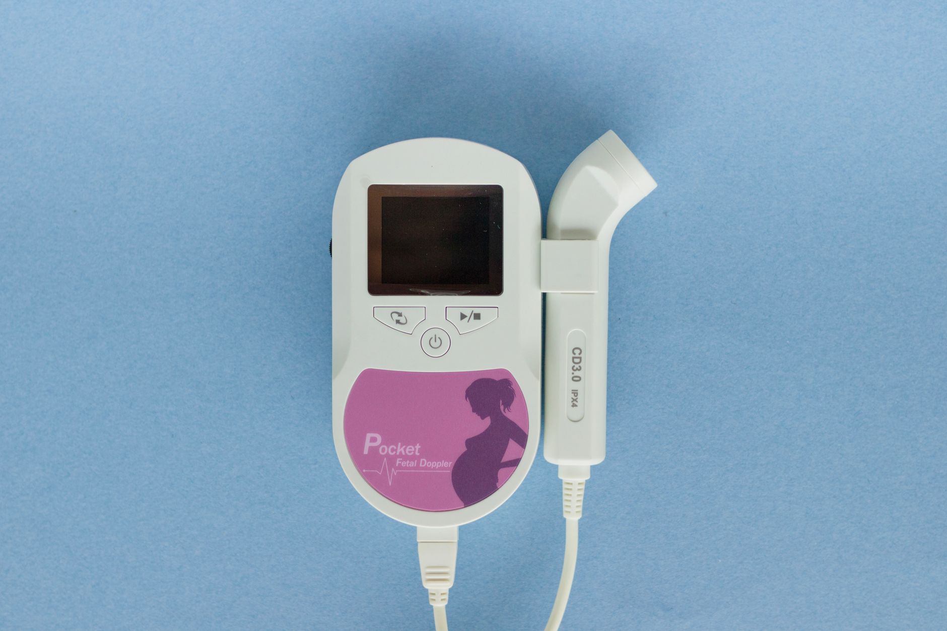
A doppler ultrasound during pregnancy safely checks your baby’s heartbeat and blood flow; it’s non‑invasive, painless, and typically performed after 10 weeks to monitor fetal health.
By Shubhra Mishra — a mom of two who turned her own confusion during pregnancy into BumpBites, a global mission to make food choices clear, safe, and stress-free for every expecting mother. 💛
Check whether any food is safe during pregnancy with the BumpBites Food Safety Checker.
Quick take: Doppler ultrasound is considered safe for most pregnancies when used according to professional guidelines. It provides valuable information about fetal heart rate and blood flow, but it should be limited to medically indicated exams—usually no more than a few times per trimester. Talk to your provider if you have concerns about frequency or any unusual symptoms.
It’s 2 a.m., you’re curled up on the couch, and the gentle “whoosh‑whoosh” of a doppler ultrasound from the clinic earlier today still echoes in your mind. You wonder: Is that sound harmless? You’re not alone. Many expecting parents ask the same question, especially after hearing the device described as “ultrasound with sound waves.” This article walks you through exactly what doppler ultrasound is, how it differs from the routine “black‑and‑white” scans, and what the latest safety data say about your baby and you.
Below, we’ll break down the science, the recommended schedule, and the practical tips you need for a calm, informed prenatal journey. You’ll also find a quick‑reference table, a myth‑busting section, and a handy FAQ that answers the most common worries. All of the information is drawn from reputable bodies such as the American College of Obstetricians and Gynecologists (ACOG), the U.K.’s National Institute for Health and Care Excellence (NICE), and the World Health Organization (WHO).
Is doppler ultrasound safe for the baby during pregnancy?
Short answer: Yes, when performed by a qualified practitioner and within the limits set by professional societies, doppler ultrasound does not pose a measurable risk to fetal development. The device uses low‑frequency sound waves (usually 2–8 MHz) that bounce off moving blood cells, creating an audible “beat” that reflects the heartbeat. Because the energy emitted is far below the thresholds that cause tissue heating or cavitation, the consensus among ACOG, the Royal College of Obstetricians and Gynaecologists (RCOG), and the WHO is that occasional doppler exams are safe.
Long‑term studies, including a 2018 systematic review of over 30 000 pregnancies, found no increase in congenital anomalies or neurodevelopmental problems linked to routine doppler use. The review emphasized that risks only appear when the ultrasound is applied for non‑medical reasons or at intensities far above diagnostic levels. In short, the “whoosh‑whoosh” you hear is a harmless by‑product of a tool that helps clinicians monitor your baby’s well‑being.
What the safety guidelines say
Both ACOG and NICE advise that doppler ultrasound should be used only when there is a clear clinical indication—such as confirming fetal heart activity, assessing blood flow in the umbilical cord, or evaluating suspected growth restriction. The guidelines also recommend keeping the thermal index (TI) and mechanical index (MI) as low as reasonably achievable. Most modern machines automatically stay within these limits, but it’s still good practice to ask your sonographer about the settings.
Doppler ultrasound pregnancy safety guidelines
Use only when medically indicated.
Limit exposure time to under 5 minutes per exam.
Maintain a safe distance—typically the transducer is placed on the abdomen, not inside the uterus.
Follow the “as low as reasonably achievable” (ALARA) principle for acoustic output.
These points are reinforced by the FDA’s 2022 guidance on diagnostic ultrasound, which notes that the thermal and mechanical indices for obstetric Doppler should remain well below the thresholds that could cause biologic effects. By staying within these limits, the technology remains a low‑risk monitoring tool throughout pregnancy.
How often can a doppler ultrasound be performed in each trimester?
There isn’t a one‑size‑fits‑all schedule, but most guidelines suggest a maximum of two to three doppler examinations per trimester for low‑risk pregnancies. In high‑risk situations—like pre‑eclampsia, fetal growth restriction, or multiple gestation—your provider may order more frequent monitoring, sometimes as often as weekly in the third trimester.
Here’s a practical breakdown:
First trimester (0‑13 weeks): Usually only one doppler scan to confirm fetal cardiac activity after the heartbeat is first seen on a standard ultrasound.
Second trimester (14‑27 weeks): If the pregnancy is uncomplicated, a doppler exam may be done once to assess umbilical artery flow and fetal well‑being.
Third trimester (28‑40 weeks): For routine care, one doppler scan is common. If there’s a concern—like decreased fetal movements—additional scans may be scheduled.
Remember, the exact number depends on your individual risk profile. Your obstetrician will tailor the schedule, balancing the need for information with the principle of minimizing unnecessary exposure.
Doppler ultrasound frequency limits during pregnancy
Professional societies set a hard limit of 30 minutes total doppler exposure per pregnancy for low‑risk patients. This limit is far above the typical cumulative exposure (often under 10 minutes) for most women. The key is to keep each individual session brief—ideally under 5 minutes—while still obtaining the needed diagnostic data.
When a high‑risk condition is identified, the cumulative limit can be adjusted, but each scan still respects the ALARA principle. In practice, the total time spent in the exam room is usually dominated by the standard B‑mode scan, with the doppler portion adding only a few minutes of focused assessment.
What are the risks of using doppler ultrasound on a pregnant mother?
While the technology is broadly safe, there are a few considerations to keep in mind. The main theoretical risks involve tissue heating and mechanical effects on cells, but diagnostic machines are designed to stay well below the thresholds that cause these effects. The most commonly reported concerns are:
Thermal effects: Excessive heating is unlikely at diagnostic levels, but prolonged exposure (over 30 minutes) could theoretically raise tissue temperature.
Mechanical effects: High‑intensity sound waves can cause tiny bubbles in fluids (cavitation), but this is not a concern with standard doppler settings.
False reassurance or anxiety: Occasionally, an abnormal doppler reading can cause unnecessary worry. It’s essential to discuss results with a qualified clinician.
If you have a condition such as a placenta previa or an active infection, your provider may postpone doppler scanning until it’s safer, because the device must be placed directly over the abdomen.
Contraindications and special considerations
Absolute contraindications are rare, but the following situations merit caution:
Known fetal anomalies that affect blood flow patterns (e.g., severe cardiac defects).
Maternal skin infections over the abdomen.
Pregnancy achieved via assisted reproductive technology where early monitoring is critical—some clinics limit doppler use to avoid unnecessary exposure.
In addition, the NHS advises that women with a history of uterine surgery should discuss any planned doppler exams with their obstetrician, as scar tissue may affect acoustic transmission.
Doppler ultrasound vs traditional ultrasound safety comparison
Traditional (B‑mode) ultrasound creates a static, gray‑scale image, while doppler adds a moving‑blood‑flow overlay and an audible heartbeat. Both use the same basic physics—high‑frequency sound waves—but doppler’s extra processing can increase acoustic output slightly. Below is a concise comparison of safety metrics across three common modalities.
Modality
Typical Frequency (MHz)
Typical Exposure Time per Exam
Thermal Index (TI) Range
Common Clinical Use
Standard B‑mode ultrasound
2‑8
5‑10 min
0.1‑0.5
Anatomical visualization, dating, growth
Doppler ultrasound
2‑8 (with added pulse‑wave)
≤5 min (recommended)
0.5‑1.0
Fetal heart rate, umbilical/placental blood flow
3‑D/4‑D ultrasound
2‑8 (plus volumetric imaging)
10‑15 min
0.5‑1.2
Detailed anatomy, parental bonding
All three modalities stay within safety margins set by the FDA and international bodies. The key difference is that doppler adds a short burst of higher‑intensity sound to capture blood flow, so the recommendation is to keep each doppler session brief. In everyday practice, the combined exposure from a standard scan plus a doppler exam rarely exceeds the safety limits.
Doppler ultrasound vs 3D ultrasound safety
Because 3‑D scans often last longer and may involve multiple angles, some guidelines advise limiting them to special occasions (e.g., showing a facial profile). Doppler, by contrast, is typically brief and targeted, making it the preferred tool for ongoing monitoring of heart rate and blood flow.
It’s also worth noting that the British Medical Ultrasound Society (BMUS) recommends reserving 3‑D imaging for specific clinical indications, whereas doppler can be incorporated into routine antenatal visits without adding significant time or thermal load.
Can doppler ultrasound detect fetal heart rate accurately?
Yes. Doppler ultrasound is the gold standard for bedside fetal heart rate (FHR) assessment when a cardiotocograph (CTG) is not available. The device detects the motion of red blood cells in the fetal heart, converting that motion into an audible “beat” and a numeric reading displayed on the screen. Accuracy is usually within ±1 bpm, comparable to electronic fetal monitoring.
In low‑risk pregnancies, doppler can reliably detect a heart rate between 110 and 160 bpm—the normal range for a healthy fetus. For high‑risk cases, such as suspected arrhythmia, a more detailed echocardiogram may be ordered, but doppler still serves as an excellent screening tool.
Benefits of fetal heart rate monitoring with doppler
Immediate reassurance when a heartbeat is heard for the first time.
Quick bedside assessment during prenatal visits.
Non‑invasive, painless, and does not require a full‑size ultrasound machine.
Limitations
While doppler is accurate for detecting the presence and basic rate of the fetal heartbeat, it cannot diagnose complex rhythm abnormalities. For that, a detailed fetal echocardiogram—often performed by a pediatric cardiologist—is required.
What does a doppler ultrasound sound like and does it affect the baby?
The “whoosh‑whoosh” you hear is the Doppler transducer translating blood flow into sound. The pitch changes with the speed of blood moving through the heart or vessels, which is why a healthy heartbeat sounds like a steady “ba‑ba‑ba.” The sound itself does not harm the baby; it is simply a by‑product of the ultrasound physics.
Many parents find the audible beat reassuring, especially during a first‑trimester scan when they hear the first heartbeat. If the sound is unsettling, you can ask the sonographer to lower the volume or mute the speaker while still visualizing the waveform on the screen.
Does the sound affect fetal development?
Research indicates that the audible component of doppler ultrasound does not influence fetal neurodevelopment. Studies measuring newborn auditory processing after prenatal exposure to doppler sounds found no differences compared with unexposed infants. The key factor remains the acoustic energy, not the audible output.
One UK cohort study published by the NHS in 2021 specifically examined auditory outcomes in children whose mothers had multiple doppler scans; the investigators reported no statistically significant effect on language milestones at age two.
Guidelines for doppler ultrasound usage in high‑risk pregnancies
High‑risk pregnancies—such as those complicated by pre‑eclampsia, intra‑uterine growth restriction (IUGR), or multiple gestations—often require more frequent doppler monitoring. The goal is to track blood flow in the umbilical artery, uterine arteries, and sometimes the middle cerebral artery (MCA) to gauge how well the placenta is delivering oxygen.
ACOG’s “Management of High‑Risk Pregnancy” guideline recommends:
Baseline doppler assessment at diagnosis of the high‑risk condition.
Serial doppler exams every 1‑2 weeks in the third trimester for IUGR or pre‑eclampsia.
Immediate doppler evaluation if the mother reports decreased fetal movements.
These more frequent scans are still within safety limits because each individual exam remains brief. The benefit of early detection of placental insufficiency outweighs the minimal theoretical risk.
Maternal blood flow assessment
Doppler can also evaluate maternal uterine artery flow, helping clinicians predict the development of pre‑eclampsia. Abnormal uterine artery waveforms early in pregnancy (high resistance index) have been linked to later hypertension. Monitoring these patterns can guide preventive strategies, such as low‑dose aspirin, as recommended by NICE.
How to interpret doppler ultrasound results for fetal wellbeing
When you receive doppler results, the sonographer will usually discuss three main parameters:
Fetal heart rate (FHR): A normal range is 110‑160 bpm. Rates outside this range may prompt further testing.
Umbilical artery (UA) Doppler: The resistance index (RI) or systolic/diastolic ratio (S/D) should decrease as pregnancy progresses. An elevated S/D ratio can signal placental insufficiency.
Middle cerebral artery (MCA) Doppler: In cases of fetal anemia or hypoxia, the MCA may show a “brain‑sparing” pattern—lower resistance indicating the fetus is redirecting blood to vital organs.
Interpretation is nuanced, and your provider will compare these values against gestational‑age‑specific reference charts. If any measurement falls outside the expected range, the next steps may include additional imaging, biophysical profiles, or early delivery planning.
Practical tip for parents
Ask your sonographer to show you the waveform on the screen and explain the numbers. A visual cue—like a smooth, low‑resistance curve—can be reassuring, and it empowers you to ask informed follow‑up questions at your next visit.
Seeing the doppler probe on the abdomen helps you understand how the exam feels.
Understanding Doppler indices: RI, PI, and S/D ratio explained
When you hear terms like “resistance index (RI)” or “pulsatility index (PI),” it can feel like medical jargon. In simple terms, these numbers describe how much resistance blood encounters as it travels through a vessel. A high RI or PI means the blood is meeting more resistance, which can be a sign that the placenta isn’t delivering oxygen as efficiently as it should.
During a normal pregnancy, the RI in the umbilical artery gradually falls because the placenta becomes more permissive to blood flow. If the RI stays high in the third trimester, clinicians may suspect growth restriction and schedule closer monitoring. The S/D ratio is another way of expressing the same concept—comparing the peak (systolic) to the trough (diastolic) of the blood‑flow waveform.
These indices are plotted on gestational‑age‑specific charts. Your provider will interpret whether your values fall within the 5th–95th percentile range, which is considered normal. If you’re ever unsure, ask for a copy of the chart; many clinics provide printable versions for patient education.
Preparing for a doppler ultrasound appointment
A doppler exam is quick, but a few simple steps can make the experience smoother. First, wear comfortable clothing that allows easy access to your abdomen—think a loose‑fitting top or a maternity shirt with a front opening. Second, stay hydrated; a full bladder can improve acoustic coupling for early‑pregnancy scans, though many doppler exams don’t require a full bladder.
Bring a list of any recent symptoms (e.g., changes in fetal movement, mild abdominal pain, or new medications) so the sonographer can tailor the scan. If you’re nervous about the “whoosh‑whoosh” sound, let the technician know ahead of time; they can lower the speaker volume or provide a quiet headset.
Finally, schedule the appointment when you’re relaxed—perhaps after a light meal and a short walk. This helps keep your heart rate steady, which can make the fetal heartbeat easier to capture.
Alternative tools: When might a home doppler be appropriate?
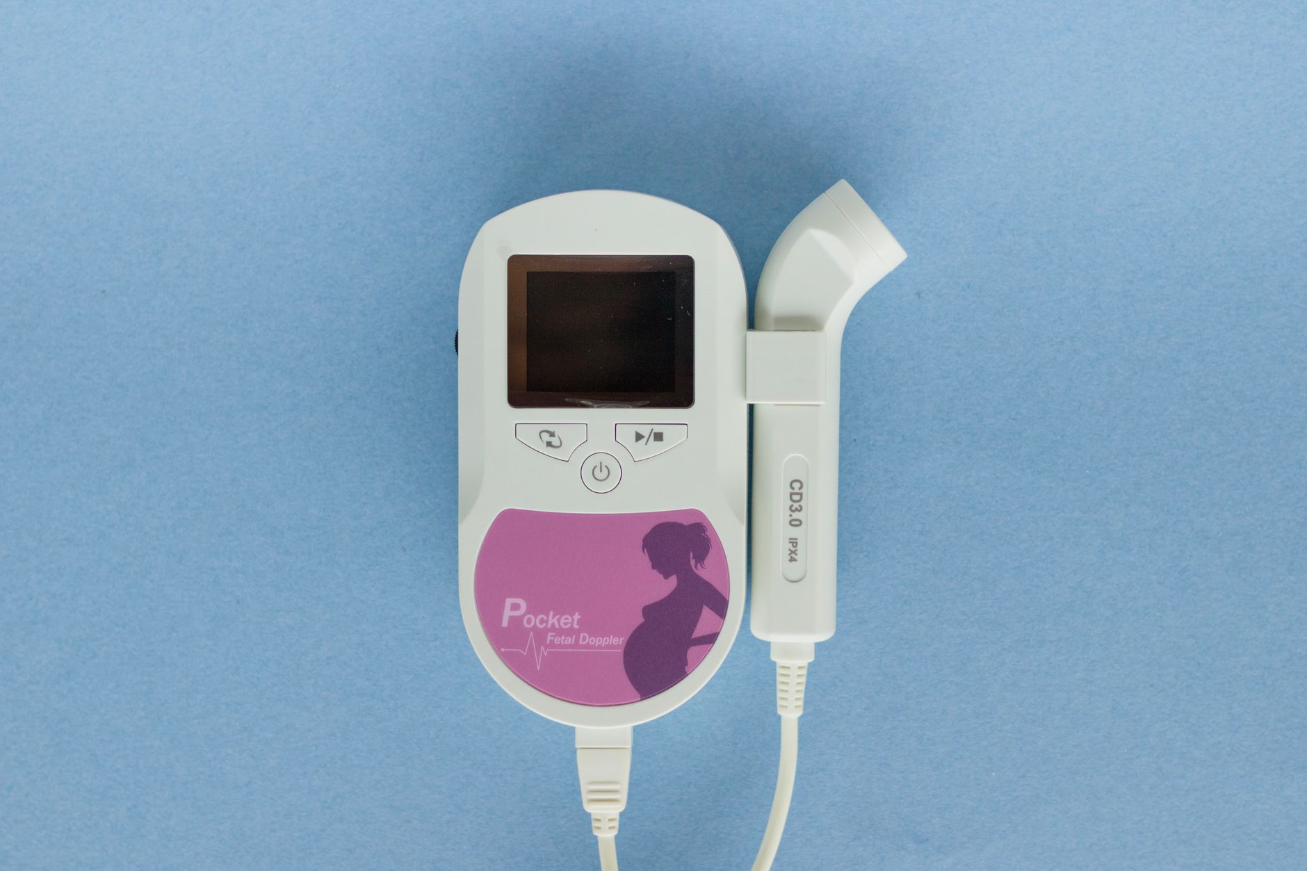
Home fetal doppler devices have become popular on online marketplaces, promising “real‑time” heartbeats for expectant parents. While they can be a fun way to bond, medical societies caution that unsupervised use carries risks. The main concerns are inaccurate readings (misreading maternal pulse as fetal) and false reassurance that may delay seeking care for genuine problems.
ACOG’s patient‑information sheet states that home dopplers should only be used after a clinician explains how to differentiate maternal and fetal signals. Even then, they recommend limiting use to a few minutes per day and never substituting professional scans. If you’re considering a home device, discuss it with your provider first to ensure you understand the limitations and proper technique.
When is doppler ultrasound safe during labor?
During active labor, a handheld doppler is often employed to monitor fetal heart rate when continuous electronic monitoring isn’t indicated. The same safety principles apply: the device emits low‑intensity sound, and the exposure is brief—usually a few minutes at a time. The American College of Nurse‑Midwives (ACNM) notes that intermittent doppler monitoring is a safe alternative to continuous CTG for low‑risk labors, provided the provider assesses the heart rate at regular intervals.
Because labor can be a stressful time, the audible beat can also reassure parents that the baby is still active. However, if the baby’s heart rate falls outside the normal range, the care team will promptly transition to continuous monitoring or additional testing.
From our medical team: Doppler ultrasound is a valuable, low‑risk tool for monitoring fetal heart rate and blood flow. Use it when your provider recommends, keep the number of scans within guideline limits, and always discuss any abnormal findings with a qualified clinician. If you ever feel uneasy during an exam, ask the technician to pause and explain what they’re seeing.
Myth vs. fact
Myth: Doppler ultrasound can cause birth defects.
Fact: Extensive research, including large cohort studies reviewed by ACOG, shows no link between diagnostic doppler use and congenital anomalies.
Myth: The audible “beat” can damage the baby’s hearing.
Fact: The sound is a by‑product of the scan and is far below the intensity that could affect fetal auditory development.
Myth: You can get unlimited doppler scans for reassurance.
Fact: While occasional scans are safe, unnecessary repeated exposure adds no benefit and may cause anxiety; follow your provider’s schedule.
Key takeaways
Doppler ultrasound is safe when used according to ACOG and NICE guidelines.
Typical limits are ≤5 minutes per exam and no more than 2‑3 scans per trimester for low‑risk pregnancies.
It reliably detects fetal heart rate (110‑160 bpm) and assesses blood flow in key vessels.
High‑risk pregnancies may need more frequent doppler monitoring, but each session remains brief.
Ask your sonographer to show you the waveform and explain any abnormal numbers.
If you notice reduced fetal movements, unusual pain, or any concerning symptoms, contact your provider promptly.
Frequently asked questions
Is doppler ultrasound safe for the baby?
Yes—when performed by a qualified professional and within recommended exposure limits, doppler ultrasound does not increase the risk of birth defects or developmental problems.
How many doppler ultrasounds can a pregnant woman have?
For low‑risk pregnancies, most guidelines advise no more than 2‑3 doppler exams per trimester, keeping each under five minutes. High‑risk cases may require more frequent monitoring as directed by your provider.
What does a doppler ultrasound sound like?
The device produces a rhythmic “whoosh‑whoosh” that mirrors the fetal heartbeat. The sound is harmless and simply reflects the movement of blood cells within the heart or vessels.
Can doppler ultrasound detect fetal heart problems?
Doppler can identify abnormal heart rates and some flow patterns, but it cannot replace a detailed fetal echocardiogram for diagnosing complex cardiac anomalies.
Are there any risks associated with doppler ultrasound?
Risks are minimal when following professional guidelines. Theoretical concerns like tissue heating are mitigated by keeping exposure time short and using low acoustic output. The main practical risk is false reassurance or anxiety from abnormal readings, which should be discussed with your clinician.
How does doppler ultrasound differ from regular ultrasound?
Standard (B‑mode) ultrasound creates static images of anatomy, while doppler adds a moving‑blood‑flow overlay and audible heartbeat. Doppler focuses on blood velocity, making it ideal for assessing fetal heart rate and placental circulation.
Can I use a home doppler device safely?
Home dopplers can be useful for bonding, but they should only be used after your provider explains how to distinguish fetal from maternal signals. Unsupervised use may lead to misinterpretation; always discuss any home‑monitoring findings with your clinician.
Is doppler ultrasound safe during labor?
Yes. Intermittent handheld doppler monitoring during low‑risk labor follows the same safety principles as prenatal scans—brief exposure, low acoustic output, and professional interpretation.
Seeing the waveform helps you understand the baby’s heartbeat in real time.
When to call your doctor
If you experience any of the following, seek medical attention promptly: sudden abdominal pain, vaginal bleeding, loss of fetal movement for more than 12 hours, persistent high fever, or a rapid rise in blood pressure (≥140/90 mmHg). Remember, this article provides general information and is not a substitute for personalized medical advice.
References
American College of Obstetricians and Gynecologists (ACOG). “Ultrasound use in pregnancy.” Practice Bulletin No. 175, 2016.
Royal College of Obstetricians and Gynaecologists (RCOG). “Guidelines for obstetric ultrasound.” 2020.
National Institute for Health and Care Excellence (NICE). “Antenatal care for uncomplicated pregnancies.” NG123, 2021.
World Health Organization (WHO). “Recommendations on antenatal care for a positive pregnancy experience.” 2016.
Food and Drug Administration (FDA). “Guidance on diagnostic ultrasound safety.” 2022.
International Society of Ultrasound in Obstetrics and Gynecology (ISUOG). “Safety of Doppler ultrasound in pregnancy.” 2019.
British Medical Ultrasound Society (BMUS). “Clinical use of Doppler ultrasound.” 2020.
Centers for Disease Control and Prevention (CDC). “Prenatal care and ultrasound safety.” 2023.
National Health Service (NHS). “Ultrasound in pregnancy: safety and outcomes.” 2021.
American College of Nurse‑Midwives (ACNM). “Intermittent fetal monitoring during labor.” Position Statement, 2020.
Not sure about the label on Doppler Ultrasound Pregnancy products?
Snap the ingredients list and SafeFilter checks every ingredient for your stage — only 3 free scans this month, then you're locked until reset. Unlimited from $7/mo or lock $50/yr through Aug 31 (5 days left).
When Shubhra Mishra was expecting her first child in 2016, she was overwhelmed by conflicting food advice — one site said yes, another said never. By the time her second baby arrived in 2019, she realized millions of mothers face the same confusion.
That sparked a five-year journey through clinical nutrition papers, cultural diets, and expert conversations — all leading to BumpBites: a calm, compassionate space where science meets everyday motherhood.
Her long-term vision is to build a global community ensuring safe, supported, and free deliveriesfor every mother — because no woman should face pregnancy alone or uninformed. 🌿
🌍 Stand with mothers, shape safer guidance
Join a small circle of experts who review BumpBites articles so expecting parents everywhere can decide with confidence.