Cord accident pregnancy can be serious. Learn what causes it, the risks to your baby, and how to reduce complications during labor and delivery.
By Shubhra Mishra — a mom of two who turned her own confusion during pregnancy into BumpBites, a global mission to make food choices clear, safe, and stress-free for every expecting mother. 💛
Check whether any food is safe during pregnancy with the BumpBites Food Safety Checker.
Quick take: An umbilical cord accident—such as a prolapse, compression, or true knot—can cause fetal distress, but most are detected early and managed with close monitoring or timely delivery. Prompt recognition, appropriate testing, and a clear delivery plan keep outcomes favorable for both baby and mother.
It’s 2 a.m., you’ve just felt a sudden, sharp cramp and a fluttering in your belly, and you’re frantically scrolling for “cord accident pregnancy.” The word alone can make your heart race. You’re not alone—many expectant parents wonder if a cord problem is happening, how to know, and what to do.
Below, we break down everything you need to know: the types of umbilical cord accidents, warning signs, how doctors diagnose them before birth, treatment options, delivery planning, and what to expect after birth. We also answer the most common follow‑up questions, from lifestyle tweaks to insurance coverage, so you can feel prepared, not panicked.
All information reflects guidance from leading bodies such as ACOG, the NHS, the CDC, and WHO. Remember, this article is for education only; your care team knows your unique situation best.
What are the signs of a cord accident during pregnancy?
A “cord accident” is an umbrella term for any event where the umbilical cord is compromised—most commonly prolapse, compression, or a true knot. While the mother may not feel the problem directly, a few subtle cues can hint that something’s amming.
Sudden change in fetal movement. A noticeable drop in kicks, or an abrupt increase in vigorous flutters, can signal fetal distress.
New or worsening abdominal pain. Sharp, localized cramping that doesn’t resolve with rest may accompany cord compression.
Vaginal bleeding or watery discharge. In the case of a cord prolapse, the membranes may rupture, leading to fluid loss.
Irregular heart‑rate patterns. On a non‑stress test (NST), a “variable deceleration”—a sudden dip and rapid return—often points to cord compression.
Most mothers won’t notice any symptom at all; that’s why routine monitoring is key. If you experience any of the above, especially a sudden lull in movement, call your provider promptly.
In addition to these signs, some women report a feeling of “pressure” in the lower abdomen, especially after a period of rapid fetal growth. While not specific, this sensation can prompt a closer look during the next prenatal visit.
How is a cord accident diagnosed before birth?
Diagnosis relies on a combination of maternal history, ultrasound imaging, and fetal monitoring.
Ultrasound
Standard obstetric ultrasound can visualize the cord’s position and detect knots or prolapse. A detailed “color Doppler” scan shows blood flow and can highlight a true knot or excessive looping.
Non‑stress test (NST) and biophysical profile (BPP)
An NST records the fetal heart rate (FHR) for 20‑40 minutes. Variable decelerations—a hallmark of cord compression—appear as abrupt drops in FHR that mirror uterine contractions. The BPP adds fetal breathing, movement, tone, and amniotic fluid volume, giving a composite score out of eight; a score ≤ 6 often prompts further evaluation.
Maternal serum markers
While not diagnostic, elevated levels of certain markers (e.g., α‑fetoprotein) can prompt a closer look for structural abnormalities, including cord issues.
In practice, a combination of a suspicious ultrasound finding and abnormal heart‑rate patterns leads clinicians to a diagnosis of a cord accident.
When an anomaly is spotted, the provider may schedule a follow‑up Doppler study within a few days to confirm the finding and assess any changes in blood flow.
Color Doppler can reveal a looping cord or true knot before birth.
Treatment options for cord accidents in the third trimester
Once a cord accident is identified after 28 weeks, the care team balances fetal safety with the risks of early delivery. Options include:
Enhanced surveillance. Daily NSTs, weekly BPPs, and twice‑weekly ultrasounds keep a close eye on fetal well‑being.
Maternal positioning. Placing the mother in a left‑lateral decubitus position can improve uterine blood flow and reduce cord compression.
Hospital admission. For high‑risk findings (e.g., persistent variable decelerations), admitting the mother for continuous fetal monitoring allows rapid response.
Planned early delivery. If the cord problem worsens or the fetus shows distress, a scheduled induction or cesarean section (C‑section) may be recommended, typically after 34 weeks when neonatal outcomes are favorable.
In most cases, careful monitoring prevents an emergency situation. However, the team remains ready to intervene promptly if the fetal heart pattern deteriorates.
Some clinicians also recommend low‑dose aspirin after 12 weeks for women at risk of placental insufficiency, as it can improve overall uteroplacental blood flow and indirectly lessen cord compression.
Can a cord accident cause stillbirth and how common is it?
Severe cord accidents can lead to stillbirth, especially if blood flow is completely cut off. According to data compiled by the CDC, umbilical cord complications account for roughly 4–6 % of stillbirths in high‑income countries. The absolute risk remains low—most pregnancies with a cord incident result in healthy live births.
Factors that raise the chance of a stillbirth include:
Prolonged cord compression with persistent variable decelerations.
True knots that tighten as the fetus grows.
Late‑term prolapse accompanied by ruptured membranes.
Early detection, close monitoring, and timely delivery dramatically reduce the likelihood of a tragic outcome.
It’s worth noting that most stillbirths are multifactorial; a cord accident rarely acts alone. Comprehensive prenatal care that includes routine growth scans and blood‑pressure checks remains the most effective prevention strategy.
Risk factors that increase chance of umbilical cord accidents
While cord accidents can happen in any pregnancy, several maternal and fetal factors raise the odds:
Excessive cord length. A longer cord (> 60 cm) can loop or knot more easily.
Polyhydramnios. Too much amniotic fluid gives the cord extra room to move.
Multiple gestation. Twins or higher-order multiples increase cord entanglement risk.
High parity. Women who have had several pregnancies have a higher chance of true knots.
Maternal diabetes. Elevated fetal activity can lead to more cord movement.
Knowing these risk factors helps providers tailor surveillance plans, but most women with risk factors still deliver without complications.
Recent ACOG guidance (2024) also highlights that maternal obesity, by altering uterine anatomy, can make cord positioning more unpredictable, underscoring the value of individualized monitoring.
What to expect during a cord accident delivery plan
When a cord accident is diagnosed, your care team will outline a delivery plan that balances safety with your preferences. Typical components include:
Location. Delivery will usually be scheduled at a hospital with full neonatal support.
Timing. If the fetus is stable, the team may aim for a gestational age that optimizes lung maturity (often ≥ 34 weeks).
Mode of delivery. Depending on the cord issue, a C‑section may be recommended. For prolapse, an emergency C‑section is often the safest route; for compression, a vaginal delivery may still be possible with close monitoring.
Team communication. Your obstetrician, midwife, anesthesiologist, and neonatologist will coordinate to ensure rapid response if fetal distress occurs.
Contingency plans. A backup operating room, blood products, and neonatal resuscitation equipment will be on standby.
Knowing these steps ahead of time can reduce anxiety and help you make informed decisions when the time comes.
Some hospitals also offer “delivery rehearsal” sessions for high‑risk pregnancies, allowing you to meet the surgical team and see the operating suite before labor begins.
How to prepare for emergency C‑section due to cord accident
Even though “emergency” sounds frightening, preparation is straightforward:
Pack a hospital bag. Include comfortable clothing, toiletries, and any items that help you relax (e.g., a music playlist).
Discuss anesthesia options. Talk with your anesthesiologist about spinal versus general anesthesia and any concerns you have.
Arrange support. Identify a partner, family member, or friend who can be present for the procedure and afterward.
Plan for newborn care. Decide whether you want skin‑to‑skin contact, breastfeeding, or a brief separation for monitoring.
Know the hospital’s policies. Some centers have specific protocols for rapid C‑sections; ask your provider what to expect.
Having these items ready means you can focus on your baby rather than logistics if an emergency arises.
Don’t forget to bring any prenatal records, a list of current medications, and a copy of your insurance card—these small details can smooth the fast‑track process.
Postnatal outcomes for babies after a cord accident
Most infants born after a cord accident have normal outcomes, especially when delivery occurs promptly. Potential short‑term issues include:
Transient tachypnea. Rapid breathing that resolves within a few hours.
Low Apgar scores. Scores of 7–8 at one minute are common but usually improve by five minutes.
Neurologic monitoring. Babies with prolonged fetal distress may undergo MRI or EEG to rule out hypoxic‑ischemic injury.
Long‑term follow‑up focuses on developmental milestones. Studies cited by the AAP indicate that, when managed appropriately, the risk of neurodevelopmental delay remains low—comparable to infants without cord complications.
Parents are encouraged to keep routine well‑baby visits and discuss any concerns about feeding, sleep, or motor development with their pediatrician.
Umbilical cord prolapse symptoms and emergency steps
A cord prolapse occurs when the cord slips through the cervix and into the birth canal, often after membranes rupture. Symptoms include a sudden gush of fluid followed by a cord‑like structure visible at the vaginal opening.
Emergency steps:
Call emergency services or your birthing center immediately.
Do not push. Avoid any maneuver that could compress the cord further.
Place the mother in a knee‑chest position. This relieves pressure on the cord by shifting the uterus upward.
Cover the cord with a sterile gauze. Keep it moist to preserve blood flow.
Prepare for rapid delivery. Most prolapses require an emergency C‑section within minutes.
After a prolapse, the obstetric team will also assess the placenta’s position and any signs of infection, as these can influence post‑delivery care.
Difference between cord compression and cord accident
“Cord compression” is a specific type of cord accident where the cord is squeezed—by the fetus’s head, a nuchal cord, or a placenta.
In contrast, “cord accident” is a broader term that includes compression, prolapse, true knots, and entanglement. All accidents involve reduced blood flow, but the mechanisms differ, influencing how clinicians monitor and intervene.
Understanding this distinction helps you interpret the language used in your medical reports and ask more precise questions at appointments.
How to monitor fetal heart rate for cord issues
Fetal heart‑rate monitoring is the frontline tool for detecting cord problems. Key patterns to watch:
Variable decelerations. Sudden drops in FHR that vary in timing and shape, often linked to cord compression.
Late decelerations. Gradual declines that begin after a contraction, indicating uteroplacental insufficiency.
Reduced baseline variability. A flat heart‑rate line can signal chronic hypoxia.
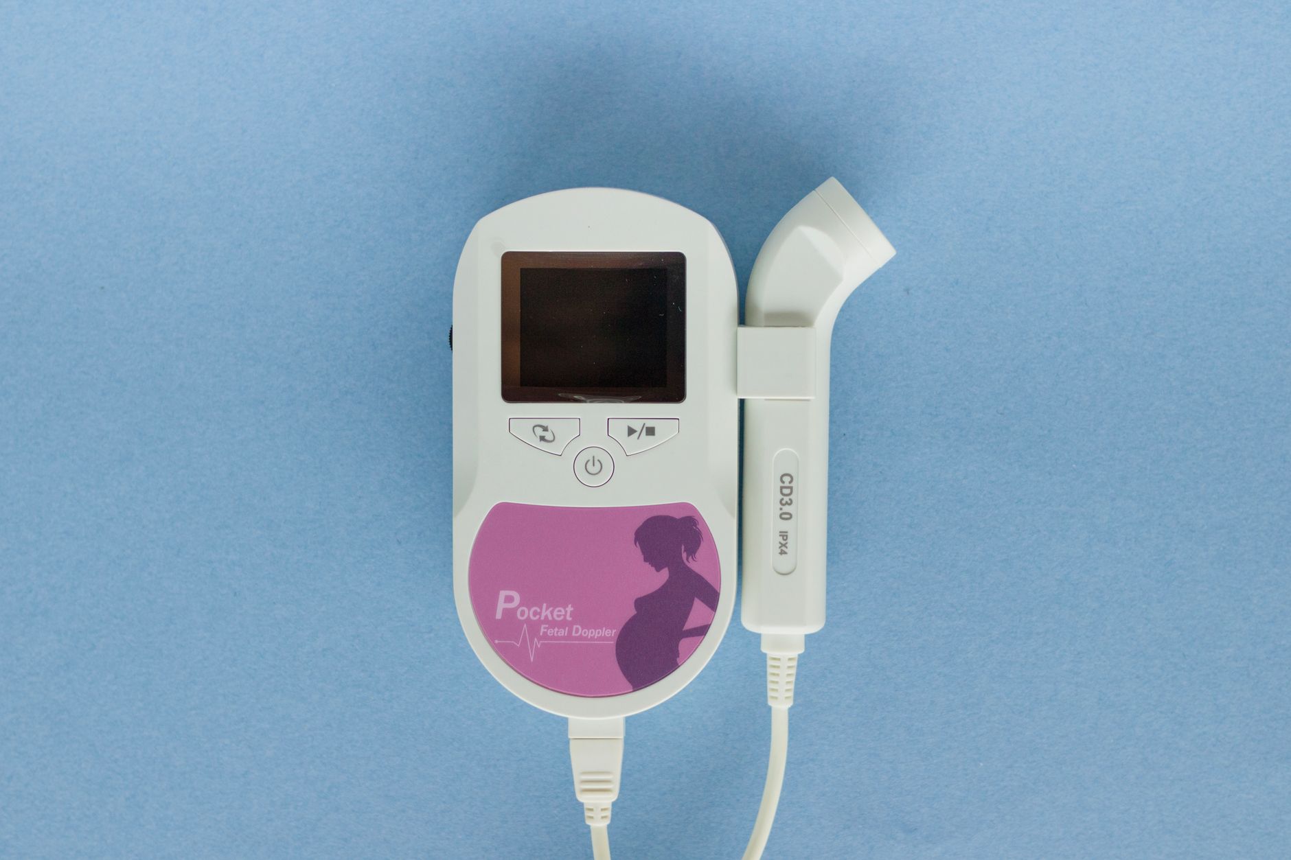
At home, some women use handheld Doppler devices, but these cannot replace professional NSTs. If you notice any abnormal pattern on a home monitor, contact your provider right away.
Professional monitoring may also include a “reactivity” test, where the fetus’s heart rate should accelerate with movements; lack of reactivity can hint at ongoing cord stress.
Best prenatal tests to detect cord problems
Several non‑invasive tests help uncover cord issues before they cause distress:
Test
What it Detects
Typical Timing
Standard obstetric ultrasound
Cord length, looping, knots
12‑20 weeks (anomaly scan)
Color Doppler ultrasound
Blood flow, true knots, prolapse
20‑24 weeks (targeted scan)
Non‑stress test (NST)
Variable decelerations, baseline variability
28 weeks onward (if risk present)
Biophysical profile (BPP)
Combined fetal activity & fluid status
28‑32 weeks (high‑risk)
Maternal serum alpha‑fetoprotein
Screen for structural anomalies
16‑18 weeks
These tests are part of routine prenatal care, but providers may schedule extra scans if a cord accident is suspected.
When a true knot is identified, the provider will often repeat Doppler imaging every 2–3 weeks to see whether the knot is tightening, a practice endorsed by the RCOG 2021 guideline.
Statistics of cord accidents in low‑risk pregnancies
Even in low‑risk pregnancies—defined by the NHS as women without chronic disease, previous complications, or multiple gestations—umbilical cord accidents still occur. A UK cohort study reported an incidence of about 1 in 500 pregnancies for any cord‑related event, with true knots present in roughly 1 % of births. While the numbers are small, they underscore the importance of universal screening during the anatomy scan.
Importantly, the same study found that over 90 % of low‑risk pregnancies with a detected knot delivered healthy infants after a planned induction or careful monitoring.
What is the role of amniotic fluid in cord accidents?
Amniotic fluid acts as a cushion, allowing the cord to move freely. Both too little (oligohydramnios) and too much (polyhydramnios) can predispose to accidents:
Polyhydramnios. Excess fluid gives the cord more space to loop, increasing the risk of knots and prolapse.
Oligohydramnios. Low fluid volume can cause the cord to become compressed against the uterine wall or placenta.
Regular ultrasound assessments of fluid volume help clinicians anticipate and mitigate these risks.
Therapeutic amnio‑infusion—a controlled infusion of saline into the amniotic cavity—is sometimes used in severe cases of oligohydramnios to reduce cord compression, though it remains a relatively rare intervention.
Can lifestyle changes reduce risk of cord accidents?
While many cord accidents are anatomical, certain lifestyle choices can lower the odds of complications that exacerbate them:
Quit smoking. Smoking narrows blood vessels, worsening any cord compression.
Maintain a balanced diet. Proper nutrition reduces the chance of diabetes, which can increase fetal activity and cord movement.
Gentle exercise. Walking and prenatal yoga promote circulation without stressing the uterus.
These measures are recommended by the WHO and ACOG for overall pregnancy health and may indirectly protect against cord accidents.
In addition, maintaining a healthy weight (BMI < 30) can lessen the mechanical pressure on the uterus, which some studies suggest may reduce cord entanglement in late pregnancy.
Insurance coverage for emergency delivery due to cord accident
Most U.S. health plans—including Medicare, Medicaid, and private insurers—cover emergency C‑sections when medically indicated, such as a cord prolapse or severe compression. In the UK, NHS care is provided at no direct cost to the patient, covering emergency deliveries and neonatal intensive care if needed.
Before pregnancy, review your policy’s “in‑network hospital” and “emergency obstetric” clauses. If you have a high‑deductible plan, ask your insurer whether pre‑authorization is required for an unscheduled C‑section, and keep a copy of your prenatal records handy.
Some insurers also offer “maternity add‑ons” that cover additional monitoring visits; these can be valuable if you’re flagged as high‑risk for cord issues.
Creating a calm environment can help you manage stress while awaiting test results.
From our medical team: Umbilical cord accidents are unsettling, but with modern monitoring and timely delivery, most babies thrive. If you notice any change in fetal movement or receive abnormal monitoring results, contact your provider right away. Your care team will tailor a plan that balances safety with your birth preferences.
How does maternal blood pressure affect cord accidents?
Elevated maternal blood pressure—whether from chronic hypertension or pregnancy‑induced conditions like pre‑eclampsia—can influence uteroplacental blood flow. Reduced perfusion may make the cord more vulnerable to compression, especially during contractions.
ACOG’s 2023 guideline recommends tighter blood‑pressure control after 20 weeks for women with gestational hypertension, because stable maternal hemodynamics lower the chance of variable decelerations that signal cord stress.
Women with well‑controlled blood pressure typically undergo more frequent NSTs, allowing clinicians to catch any early signs of cord compromise before they become critical.
Can early‑pregnancy ultrasound detect cord problems?
Standard first‑trimester scans focus on confirming viability and dating, but they can also give an early glimpse of cord length and placement. While true knots are rare this early, a markedly long cord (> 70 cm) can already be visualized and flagged for later surveillance.
The NHS recommends a detailed anatomy scan between 18‑22 weeks, which is the optimal window to assess cord looping, nuchal cords, and potential prolapse. Detecting an issue at this stage provides ample time for a tailored monitoring schedule.
In cases where a cord anomaly is seen early, a follow‑up Doppler study at 24‑28 weeks is often scheduled to track any changes in blood flow or cord configuration.
Nutrition and supplements that support cord health
While no food can change cord length, certain nutrients promote overall placental and fetal vascular health, indirectly supporting the cord’s function.
Omega‑3 fatty acids. Found in fortified eggs, low‑mercury fish, and flaxseed, they help maintain healthy blood vessels, as highlighted by the FDA’s pregnancy nutrition label.
Folate‑rich foods. Leafy greens, legumes, and fortified cereals reduce the risk of neural‑tube defects and support proper fetal circulation.
Vitamin D. Adequate levels, achieved through sunlight exposure and fortified dairy, are linked to lower rates of pre‑eclampsia, which in turn steadies uterine blood flow.
Always discuss supplement use with your provider, especially if you have a history of kidney stones or are on anticoagulant therapy, as excessive vitamin K can affect clotting during delivery.
Myth vs. fact
Myth: A cord accident always leads to a stillbirth.
Fact: While severe cord compression can cause fetal distress, most cord accidents are identified early and managed with monitoring or delivery, resulting in healthy outcomes.
Myth: You can feel a cord accident as a sharp pain.
Fact: Most mothers do not feel the cord itself; the warning signs are usually changes in fetal movement or abnormal heart‑rate patterns on monitoring.
Myth: Once a cord accident is diagnosed, you must have an immediate C‑section.
Fact: Management depends on gestational age, fetal status, and the type of accident; many cases are monitored closely until a safe delivery window is reached.
Key takeaways
Umbilical cord accidents include prolapse, compression, and true knots; most are detected via ultrasound or fetal monitoring.
Sudden changes in fetal movement or variable decelerations on NST are key warning signs.
Enhanced surveillance, maternal positioning, and timely delivery keep outcomes favorable.
Risk factors include long cords, polyhydramnios, multiple gestations, and smoking.
Emergency C‑section preparation involves packing a hospital bag, discussing anesthesia, and arranging support.
Post‑delivery, most babies do well; long‑term neurodevelopmental issues are rare when care is prompt.
Maternal blood‑pressure control and adequate nutrition further reduce the chance of cord‑related complications.
Frequently asked questions
What does a cord accident feel like for the mother?
Most women do not feel the cord itself; the most common sensation is a sudden change in fetal movement—either a lull or an increase in flutters.
How quickly can a cord accident lead to fetal distress?
Depending on the type, distress can develop within minutes (as with prolapse) or over hours (as with a tightening knot); continuous monitoring helps catch changes early.
Can a cord accident be prevented during pregnancy?
While you cannot control cord length, staying hydrated, avoiding smoking, and managing diabetes reduce related risks and support overall uterine health.
What are the emergency procedures if a cord accident occurs during labor?
Immediate steps include calling the care team, placing the mother in a knee‑chest position, keeping the cord moist, and preparing for a rapid C‑section.
Is a cord accident more common in first‑time mothers?
True knots are slightly more frequent in multiparous women, but overall cord accidents affect both first‑time and experienced mothers alike.
What are the long‑term effects on a baby after a cord accident?
When managed promptly, most infants have normal growth and development; rare cases of severe hypoxia may require early intervention, but long‑term outcomes are generally favorable.
What should I do if my home Doppler shows a variable deceleration?
Contact your provider right away; they will likely schedule an urgent NST or BPP to assess the fetus’s well‑being and determine whether additional monitoring or delivery is needed.
Are there any lasting health considerations for the mother after a cord accident?
Most mothers recover without complications. However, if the accident was linked to a condition like pre‑eclampsia or severe anemia, follow‑up appointments may be recommended to monitor blood pressure and iron levels postpartum.
When to call your doctor
Seek immediate medical attention if you notice any of the following: a sudden decrease or increase in fetal movement, vaginal bleeding or fluid loss, persistent variable decelerations on a home monitor, or any visible cord protruding from the vagina. This article is for educational purposes only and does not replace personalized medical advice.
References
American College of Obstetricians and Gynecologists (ACOG). “Management of Umbilical Cord Complications.” Clinical Guidance, 2023.
American College of Obstetricians and Gynecologists (ACOG). “Blood Pressure Management in Pregnancy.” Obstetrics & Gynecology, 2024.
National Health Service (NHS). “Umbilical Cord Problems.” Patient Information, 2022.
Centers for Disease Control and Prevention (CDC). “Stillbirth and Cord Abnormalities.” Vital Statistics Report, 2021.
World Health Organization (WHO). “Antenatal Care Guidelines.” Global Recommendations, 2022.
American Academy of Pediatrics (AAP). “Neonatal Outcomes After Umbilical Cord Compression.” Pediatrics, 2020.
Royal College of Obstetricians and Gynaecologists (RCOG). “Ultrasound Assessment of the Umbilical Cord.” Green‑top Guideline, 2021.
National Institute for Health and Care Excellence (NICE). “Fetal Monitoring in Pregnancy.” Clinical Guideline CG190, 2023.
U.S. Department of Health and Human Services. “Insurance Coverage for Emergency Obstetric Care.” Policy Brief, 2022.
Food and Drug Administration (FDA). “Pregnancy and Lactation Labeling Rule (PLLR).” Guidance for Industry, 2020.
World Health Organization (WHO). “Nutrition for Women During Pregnancy.” Fact Sheet, 2021.
American College of Obstetricians and Gynecologists (ACOG). “Gestational Hypertension and Preeclampsia.” Practice Bulletin, 2023.
Not sure about the label on Cord Accident Pregnancy products?
Snap the ingredients list and SafeFilter checks every ingredient for your stage — only 3 free scans this month, then you're locked until reset. Unlimited from $7/mo or lock $50/yr through Aug 31 (5 days left).
When Shubhra Mishra was expecting her first child in 2016, she was overwhelmed by conflicting food advice — one site said yes, another said never. By the time her second baby arrived in 2019, she realized millions of mothers face the same confusion.
That sparked a five-year journey through clinical nutrition papers, cultural diets, and expert conversations — all leading to BumpBites: a calm, compassionate space where science meets everyday motherhood.
Her long-term vision is to build a global community ensuring safe, supported, and free deliveriesfor every mother — because no woman should face pregnancy alone or uninformed. 🌿
🌍 Stand with mothers, shape safer guidance
Join a small circle of experts who review BumpBites articles so expecting parents everywhere can decide with confidence.