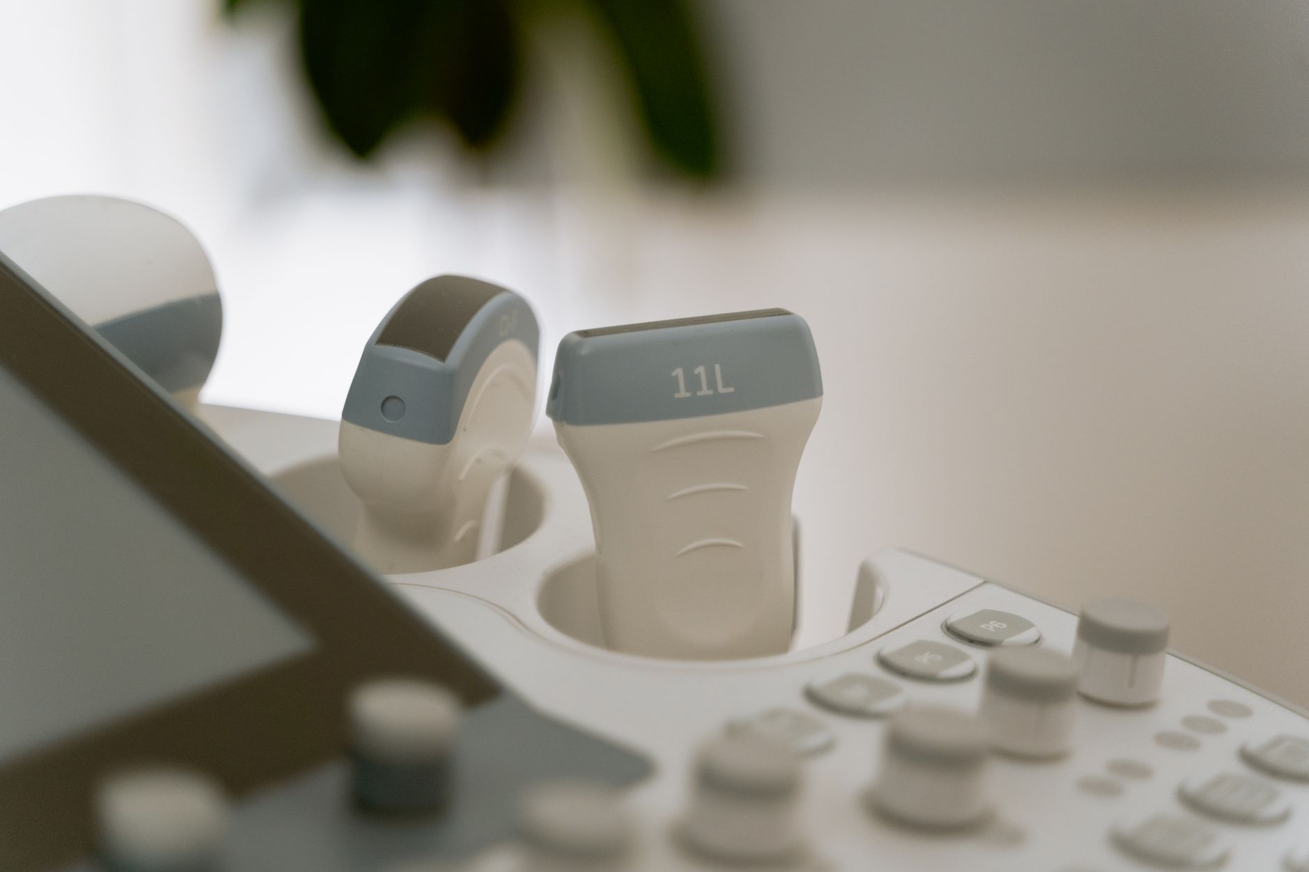
Cordocentesis is a prenatal test to diagnose fetal conditions. Learn when it’s recommended, risks like miscarriage, and how it compares to amniocentesis or CVS.
By Shubhra Mishra — a mom of two who turned her own confusion during pregnancy into BumpBites, a global mission to make food choices clear, safe, and stress-free for every expecting mother. 💛
Check whether any food is safe during pregnancy with the BumpBites Food Safety Checker.
Quick take: Cordocentesis, also called percutaneous umbilical blood sampling, is an invasive test that directly samples fetal blood from the umbilical cord. It is most often used after the second trimester to diagnose genetic disorders, fetal anemia, or infections, and it carries a slightly higher risk of fetal loss than amniocentesis. Your provider will weigh the benefits against the small risk before recommending it.
It’s 2 a.m., you’ve just felt a flutter in your belly and the worry that something might be wrong pops up again. You wonder whether the “cord blood test” you read about could give you the answers you need. You’re not alone—many expecting parents ask, “Is cordocentesis safe? When should it be done? What will it tell us?” In this guide we break down everything you need to know about cordocentesis, from what the procedure looks like to how accurate it is, and what you can expect before, during, and after the test.
We’ll walk through the definition, timing, step‑by‑step process, risks, and how it compares with other prenatal tests like amniocentesis and chorionic villus sampling (CVS). You’ll also find practical tips on preparation, recovery, cost, and insurance, plus a clear list of red‑flag symptoms that require immediate medical attention. By the end, you’ll have a realistic picture of what this specialized test involves and when it might be the right choice for you.
What is cordocentesis and how does it differ from amniocentesis?
Cordocentesis—formally known as percutaneous umbilical blood sampling (PUBS)—involves inserting a thin needle through the mother’s abdomen, through the uterus, and into the fetal umbilical cord to obtain a small amount of fetal blood. The sample can be analyzed for chromosomes, genetic mutations, infections, and fetal anemia.
Amniocentesis, by contrast, samples the amniotic fluid that surrounds the baby. While amniotic fluid contains fetal cells, cordocentesis gives a direct blood sample, which often provides clearer results for certain conditions, especially when the fetus is already in the second or third trimester.
Key differences:
Timing: Amniocentesis is usually performed between 15‑20 weeks; cordocentesis is typically done after 18 weeks, often later.
Sample type: Amniotic fluid vs. fetal blood.
Risk profile: Cordocentesis carries a slightly higher risk of fetal loss (about 1‑2 % according to ACOG) compared with amniocentesis (≈0.5‑1 %).
Diagnostic yield: Cordocentesis can diagnose infections (e.g., cytomegalovirus) and anemia more directly than amniocentesis.
Both procedures require ultrasound guidance, sterile technique, and a skilled maternal‑fetal medicine specialist. The choice depends on what your provider needs to find out and how far along you are. Because cord blood reflects the fetus’s circulating environment, it can reveal metabolic disorders that may be missed in amniotic fluid, a nuance highlighted in the ACOG Practice Bulletin on invasive prenatal diagnosis.
During cordocentesis, a real‑time ultrasound guides the needle to the umbilical cord.
Cordocentesis procedure steps and what to expect during the test
The procedure usually takes 30‑45 minutes, including preparation and post‑procedure monitoring.
1. Pre‑procedure preparation
Confirm fasting status (usually no food for 2‑4 hours).
Review your medical history, medications, and any allergies.
Obtain informed consent after discussing benefits, risks, and alternatives.
2. Positioning and anesthesia
You’ll lie on a comfortable exam table, often with a slight tilt to ease access to the uterus. A local anesthetic (e.g., lidocaine) is injected at the entry site to numb the skin and abdominal wall.
3. Ultrasound guidance
Using a high‑frequency transducer, the specialist identifies a straight segment of the umbilical cord away from the placenta and fetus. The needle, typically 22‑ or 24‑gauge, is then advanced under continuous ultrasound visualization.
4. Sampling fetal blood
Once the needle tip is confirmed within the cord vessel, a small amount (0.5‑1 mL) of fetal blood is drawn into a syringe. The sample is immediately sent to the laboratory for rapid chromosome analysis, PCR testing for infections, or hematology studies.
5. Needle removal and post‑procedure monitoring
The needle is withdrawn, a small bandage applied, and you’ll be observed for 30‑60 minutes for any bleeding or uterine cramping. A follow‑up ultrasound may be performed to ensure the placenta and fetus are stable.
Most women report mild discomfort from the needle entry and brief uterine soreness afterward—similar to a deep blood draw. The entire experience is designed to be as quick and low‑stress as possible. If you feel any sudden sharp pain during needle insertion, the team will pause and reassess the positioning before continuing.
Risks and complications associated with cordocentesis
Every invasive prenatal test carries some risk, and cordocentesis is no exception. The most commonly reported complications, as outlined by the American College of Obstetricians and Gynecologists (ACOG) and the Royal College of Obstetricians and Gynaecologists (RCOG), include:
Fetal loss: Approximately 1‑2 % risk of miscarriage or stillbirth, higher than amniocentesis.
Bleeding: Maternal or fetal bleeding at the puncture site, usually minor but occasionally requiring intervention.
Infection: Rare (<0.1 %) but possible; prophylactic antibiotics are sometimes given.
Placental abruption: Separation of placenta from uterine wall, a serious emergency.
Uterine contractions: May be triggered by the procedure; tocolytics (medication to relax the uterus) are often administered prophylactically.
Fetal arrhythmia: Temporary heart rhythm changes, usually self‑limited.
Because the needle traverses the uterine wall and directly enters the cord, the procedure demands precise ultrasound guidance and an experienced operator to minimize these risks. Your provider will discuss your individual risk factors—such as a history of preterm labor, placenta previa, or multiple gestation—before proceeding. In many centers, a dedicated fetal‑medicine nurse monitors you for at least an hour after the test to catch any early signs of complication.
When is cordocentesis recommended during pregnancy?
Cordocentesis is not a routine screening test. It is typically reserved for specific clinical scenarios where other, less‑invasive methods cannot provide the needed information.
Fetal anemia assessment: When ultrasound shows signs of anemia (e.g., increased peak systolic velocity in the middle cerebral artery), cordocentesis can directly measure hemoglobin levels.
Infection diagnosis: Suspected congenital infections such as cytomegalovirus (CMV) or toxoplasmosis, especially after a positive maternal serology.
Genetic confirmation after abnormal screening: If non‑invasive prenatal testing (NIPT) or amniocentesis yields an uncertain result, cordocentesis can provide a definitive diagnosis.
Therapeutic monitoring: In cases of fetal therapy (e.g., intra‑uterine transfusion for severe anemia), cordocentesis is used before and after treatment to assess response.
Guidelines from ACOG recommend performing cordocentesis after 18 weeks gestation, when the umbilical cord is large enough to safely accommodate the needle. In rare circumstances, it can be done earlier, but the technical difficulty and risk increase sharply before 16 weeks. Multidisciplinary teams—including maternal‑fetal medicine physicians, genetic counselors, and neonatologists—usually review each case to ensure the benefit outweighs the risk.
How accurate is cordocentesis for diagnosing fetal genetic disorders?
Because cordocentesis obtains a direct blood sample, its diagnostic accuracy for chromosomal abnormalities and single‑gene disorders is very high—often approaching 99 % concordance with post‑natal testing. The American College of Medical Genetics (ACMG) notes that the specificity for detecting trisomies (e.g., Down syndrome) and large chromosomal deletions is comparable to that of amniocentesis, while the sensitivity for metabolic disorders can be superior because fetal blood reflects the lived environment.
For infectious agents, cordocentesis allows PCR testing of the fetal blood, which is more reliable than amniotic fluid PCR when the pathogen preferentially resides in blood cells. However, the overall utility depends on the laboratory’s expertise and the timing of the infection. Advanced laboratories often use next‑generation sequencing to increase detection rates for rare genetic conditions, a practice endorsed by the FDA for certain high‑risk pregnancies.
It’s worth noting that a small percentage of results may be “inconclusive” due to sample contamination or insufficient fetal cells. In such cases, your provider may repeat the test or recommend an alternative method such as amniocentesis or a repeat NIPT.
Cordocentesis vs chorionic villus sampling: which is better?
Both cordocentesis and chorionic villus sampling (CVS) are invasive diagnostic tools, but they differ in timing, sample type, and risk profile. Below is a concise comparison:
Feature
Cordocentesis
Chorionic Villus Sampling (CVS)
Typical timing
18 weeks + (second/third trimester)
10‑13 weeks (first trimester)
Sample source
Fetal blood from umbilical cord
Placental villi (fetal tissue)
Primary uses
Genetic confirmation, anemia, infections
Early detection of chromosomal abnormalities
Risk of miscarriage
≈1‑2 % (higher)
≈0.5‑1 % (lower)
Risk of limb‑defect
None
Very rare (<0.1 %)
Need for ultrasound guidance
Yes, real‑time
Yes, for placental location
If you need an early diagnosis (before 14 weeks), CVS is the preferred option. Cordocentesis shines later in pregnancy, especially when evaluating fetal anemia or infections that cannot be assessed via CVS. Your personal values—such as how early you want information and how you weigh procedural risk—play a central role in the decision, and your care team will help you explore those preferences.
What is the recovery time after a cordocentesis procedure?
Most patients are discharged after a short observation period (30‑60 minutes). Recovery advice typically includes:
Rest at home for the remainder of the day; avoid strenuous activity or heavy lifting for 24‑48 hours.
Stay hydrated and monitor for any vaginal bleeding, fluid leakage, or severe abdominal pain.
Take prescribed analgesics (usually acetaminophen) if you experience mild cramping.
Schedule a follow‑up ultrasound within a week to confirm fetal well‑being.
Women who receive tocolytics may feel a slight drowsiness, but these medications are short‑acting. Most return to normal daily routines within a couple of days, though some choose to take it easy for a full week. Emotionally, it’s common to feel a mix of relief and lingering anxiety; many parents find it helpful to keep a brief journal of symptoms and thoughts during the first 48 hours.
Can cordocentesis be performed in the first trimester?
Technically, a needle can be inserted into the umbilical cord as early as 12 weeks, but the procedure is rarely performed before 16 weeks because the cord vessels are very small and the risk of fetal injury rises sharply. The ACOG Practice Bulletin on invasive prenatal diagnosis advises against first‑trimester cordocentesis except in exceptional circumstances, such as a life‑threatening fetal infection that cannot wait for later testing.
When early diagnosis is essential, clinicians usually opt for CVS (10‑13 weeks) or, if the concern is an infection, they may rely on maternal serology and amniocentesis (after 15 weeks) instead of a risky early cordocentesis. In those rare early‑trimester cases, a multidisciplinary review—including a neonatologist and infectious‑disease specialist—is required to justify the approach.
Take it easy after the test—gentle rest helps reduce uterine irritation.
Cordocentesis success rate statistics
Success is defined as obtaining a sufficient, uncontaminated fetal blood sample for definitive laboratory analysis. Large‑scale data from the US and UK suggest a technical success rate of 95‑98 % when performed by experienced fetal‑medicine teams. Failure is most often due to poor cord visualization or maternal obesity limiting ultrasound clarity.
When the procedure is successful, diagnostic yield for conditions such as fetal anemia or CMV infection exceeds 90 %, according to the National Institute for Health and Care Excellence (NICE) guidelines on invasive testing. Centers that employ 3‑D ultrasound or color Doppler tend to report slightly higher success rates, highlighting the importance of advanced imaging technology.
Cordocentesis fetal blood sampling guidelines
Professional bodies provide clear recommendations to standardize the test:
Needle size: 22‑ or 24‑gauge to balance sample volume with minimal trauma.
Sample volume: 0.5‑1 mL is sufficient for most genetic, hematologic, and infectious assays.
Anticoagulant: EDTA tubes are preferred for DNA analysis; heparin may be used for certain metabolic tests.
Timing of analysis: Results for chromosomal microarray or PCR are typically available within 7‑10 days.
Operator experience: ACOG recommends that the physician have performed at least 50 invasive procedures, with a minimum of 10 cordocenteses, to maintain proficiency.
In addition, the FDA requires laboratories performing fetal blood genetic testing to adhere to CLIA certification standards, ensuring consistency and reliability across facilities.
Cordocentesis cost and insurance coverage
In the United States, the out‑of‑pocket cost for cordocentesis can range from $1,500 to $3,000, depending on facility fees, laboratory charges, and whether a genetic counselor is involved. Many private insurers cover the procedure when it is deemed medically necessary—most often after a documented abnormal screening or a suspected fetal anemia.
In the United Kingdom, cordocentesis is provided through the NHS free of charge for qualifying indications, though there may be a short waiting period due to limited specialist availability. Public‑funded health systems in Canada and Australia similarly cover the test under specialist referral. If you have a high‑deductible plan, ask your provider’s billing office about pre‑authorization and possible financial assistance programs.
Cordocentesis contraindications and who cannot have it
Not everyone is a candidate for cordocentesis. Absolute contraindications include:
Placenta previa or low‑lying placenta that blocks safe needle access.
Multiple gestations where separating a single cord is technically unsafe.
Active maternal infection that could be transmitted to the fetus via the needle.
Severe maternal coagulopathy (e.g., uncontrolled thrombocytopenia).
Relative contraindications—situations that increase risk but may be considered on a case‑by‑case basis—include maternal obesity, prior uterine surgery, and a history of preterm labor. In these scenarios, a thorough risk‑benefit conversation is essential, and some centers may opt for alternative testing methods such as amniocentesis or repeat NIPT.
Cordocentesis fetal anemia diagnosis
Fetal anemia is most commonly associated with red‑cell alloimmunization (Rh disease) or parvovirus B19 infection. Cordocentesis allows direct measurement of fetal hemoglobin and reticulocyte count, providing a definitive diagnosis. The procedure also guides therapeutic decisions such as intra‑uterine transfusion.
According to the ACOG Practice Bulletin on fetal anemia, a hemoglobin level < 0.8 times the gestational age‑adjusted median or a markedly elevated middle cerebral artery peak systolic velocity on Doppler ultrasound usually prompts cordocentesis for confirmation. When anemia is confirmed, intra‑uterine transfusion can be performed in the same session, dramatically improving survival rates.
Cordocentesis ultrasound guidance techniques
High‑resolution transabdominal ultrasound is the standard, but some centers supplement it with transvaginal or 3‑D ultrasound to improve visualization of the cord insertion point. Key technical tips include:
Choosing a straight, non‑twisting segment of the cord at least 2 cm away from the placenta.
Using color Doppler to confirm arterial flow before needle insertion.
Applying gentle maternal breath‑holding to minimize fetal movement during needle entry.
Real‑time image recording for quality assurance and medico‑legal documentation.
Advanced centers may employ fetal MRI guidance in complex cases, though this is not routine. Ongoing training programs, such as those endorsed by the Society for Maternal‑Fetal Medicine (SMFM), ensure that new fellows achieve competency before performing independent procedures.
Cordocentesis vs amniocentesis safety comparison
Both procedures share common safety concerns—maternal discomfort, infection, and fetal loss—but the relative rates differ:
Fetal loss: Cordocentesis 1‑2 % vs. amniocentesis 0.5‑1 % (ACOG).
Maternal bleeding: Slightly higher with cordocentesis due to deeper uterine traversal.
Infection risk: Comparable, both <0.1 % with sterile technique.
Uterine irritability: More frequent after cordocentesis; prophylactic tocolysis is often given.
Overall, the absolute risk difference is small, but your provider will discuss these nuances in the context of your specific indication and gestational age. Some families prioritize the higher diagnostic yield of cordocentesis for anemia, while others feel more comfortable with the slightly lower risk profile of amniocentesis. Shared decision‑making tools, such as those developed by the AAP, can help clarify personal values.
From our medical team: Cordocentesis is a valuable diagnostic tool when the situation truly calls for it. The key is balancing the need for precise fetal information against the modest increase in procedural risk. If you’re considering this test, ask your provider about the exact reason for recommendation, the success rate at your center, and what monitoring will look like afterward. Most families who undergo cordocentesis find the clarity it provides outweighs the brief discomfort.
Non‑invasive alternatives: cell‑free DNA testing
Before opting for an invasive test, many parents explore non‑invasive prenatal testing (NIPT) that analyzes cell‑free fetal DNA circulating in the mother’s blood. NIPT can screen for common trisomies, sex chromosome anomalies, and some microdeletions with a detection rate above 99 % for Down syndrome, according to the FDA‑approved assays.
While NIPT is highly sensitive, it remains a screening tool—not a diagnostic test. Positive NIPT results often lead to confirmatory invasive testing, such as amniocentesis or cordocentesis, especially when the condition being screened for cannot be fully evaluated without fetal blood (e.g., fetal anemia). Discuss with your genetic counselor whether NIPT alone meets your informational needs or whether an invasive test is still warranted.
Psychological support and decision‑making
Choosing to undergo cordocentesis can feel overwhelming. Studies published by the NHS and ACOG show that parents who receive structured counseling report lower anxiety and higher satisfaction with their decision. A typical counseling session includes a clear explanation of why the test is recommended, an overview of alternatives, and a discussion of personal values such as timing of information and tolerance for procedural risk.
Many clinics offer access to a perinatal psychologist or social worker who can help you process emotions, especially if you’re dealing with a previous loss or a high‑risk pregnancy. Keeping a list of questions—like “What will happen if the test is inconclusive?” or “How will the results affect our birth plan?”—can make the appointment more productive and ensure you leave feeling informed.
Myth vs. fact
Myth: Cordocentesis is always riskier than any other prenatal test.
Fact: While the risk of fetal loss is slightly higher than amniocentesis, it remains under 2 % when performed by experienced clinicians, and the diagnostic benefit can be decisive for certain conditions.
Myth: The test can be done safely at any stage of pregnancy.
Fact: Cordocentesis is generally performed after 18 weeks; early‑trimester attempts carry higher technical difficulty and increased risk.
Myth: You’ll feel severe pain during the procedure.
Fact: Local anesthesia numbs the entry site, and most women describe the sensation as a brief pressure rather than sharp pain.
Key takeaways
Cordocentesis samples fetal blood directly, offering high‑accuracy diagnosis for anemia, infections, and many genetic conditions.
The procedure is usually done after 18 weeks and carries a 1‑2 % risk of fetal loss.
Ultrasound guidance, a skilled operator, and proper prenatal counseling are essential for safety.
Insurance often covers cordocentesis when medically indicated, but out‑of‑pocket costs can vary.
Recovery is quick—most moms rest at home for 24‑48 hours and resume normal activity within a week.
Non‑invasive options like cell‑free DNA testing can be a first step, but definitive answers for anemia or infection still require cord blood sampling.
Frequently asked questions
How long does it take to get results from cordocentesis?
Most genetic and chromosomal results are available within 7‑10 days, while infectious disease PCR can be returned in 3‑5 days; your provider will give you a specific timeline based on the tests ordered.
Is cordocentesa safe for the baby?
When performed by an experienced fetal‑medicine specialist, the overall risk of fetal loss is about 1‑2 %, which is low but higher than amniocentesis; the benefit often outweighs this small risk for critical diagnoses.
What are the signs of complications after cordocentesis?
Watch for heavy vaginal bleeding, sudden severe abdominal pain, fluid leakage from the vagina, fever, or a rapid increase in uterine contractions; any of these warrant immediate medical attention.
Can cordocentesis detect Down syndrome?
Yes—by analyzing fetal chromosomes from the blood sample, cordocentesis can confirm trisomy 21 (Down syndrome) with accuracy comparable to amniocentesis.
How many weeks pregnant can you have cordocentesis?
The test is typically performed after 18 weeks, though it can be safely done up to term; performing it before 16 weeks is rarely recommended due to technical challenges.
Is cordocentesis painful for the mother?
Local anesthesia numbs the skin and abdominal wall, so most mothers report only mild pressure or brief discomfort during needle insertion, followed by mild cramping afterward.
Will I need a special follow‑up after cordocentesis?
Yes—your provider will schedule a follow‑up ultrasound within a week to confirm fetal well‑being, and you’ll receive a detailed plan for any additional testing if the initial results are inconclusive.
Can I have cordocentesis if I have a scar from a previous C‑section?
Most providers can safely perform cordocentesis in women with a prior low transverse C‑section, but they may adjust the entry point to avoid the scar tissue; a thorough ultrasound assessment determines the safest trajectory.
When to call your doctor
If you notice any of the following after cordocentesis, contact your obstetric provider or go to the nearest emergency department right away: heavy vaginal bleeding (soaking a pad in under a minute), persistent severe abdominal pain, fever higher than 100.4 °F (38 °C), fluid leaking from the vagina, or a sudden decrease in fetal movement.
This article provides general information and is not a substitute for personalized medical advice. Always discuss your specific situation with your health care provider.
References
American College of Obstetricians and Gynecologists. Practice Bulletin No. 199: Invasive Prenatal Diagnosis. 2022.
Royal College of Obstetricians and Gynaecologists. Guidelines for Invasive Prenatal Testing. 2021.
National Institute for Health and Care Excellence (NICE). Fetal Anemia and Invasive Testing. NG126. 2020.
American College of Medical Genetics and Genomics. Laboratory Standards for Fetal Blood Sampling. 2021.
Centers for Disease Control and Prevention. Cytomegalovirus in Pregnancy. Updated 2023.
World Health Organization. Recommendations on Invasive Prenatal Diagnosis. 2022.
Mayo Clinic. Cordocentesis (Percutaneous Umbilical Blood Sampling). Accessed July 2026.
National Health Service (NHS). Chorionic Villus Sampling and Cordocentesis. Updated 2023.
Society for Maternal-Fetal Medicine. Consensus Statement on Indications for Cordocentesis. 2022.
Health Insurance Portability and Accountability Act (HIPAA) compliance guidelines for prenatal testing coverage. 2023.
Food and Drug Administration. Guidance for Cell‑Free DNA Tests in Pregnancy. 2021.
American Academy of Pediatrics. Shared Decision‑Making Tools for Prenatal Testing. 2022.
Not sure about the label on Cordocentesis products?
Snap the ingredients list and SafeFilter checks every ingredient for your stage — only 3 free scans this month, then you're locked until reset. Unlimited from $7/mo or lock $50/yr through Aug 31 (5 days left).
When Shubhra Mishra was expecting her first child in 2016, she was overwhelmed by conflicting food advice — one site said yes, another said never. By the time her second baby arrived in 2019, she realized millions of mothers face the same confusion.
That sparked a five-year journey through clinical nutrition papers, cultural diets, and expert conversations — all leading to BumpBites: a calm, compassionate space where science meets everyday motherhood.
Her long-term vision is to build a global community ensuring safe, supported, and free deliveriesfor every mother — because no woman should face pregnancy alone or uninformed. 🌿
🌍 Stand with mothers, shape safer guidance
Join a small circle of experts who review BumpBites articles so expecting parents everywhere can decide with confidence.