Amniotic fluid monitoring is performed every 1–2 weeks after the 28th week; abnormal levels trigger interventions such as surveillance, medication, or delivery.
By Shubhra Mishra — a mom of two who turned her own confusion during pregnancy into BumpBites, a global mission to make food choices clear, safe, and stress-free for every expecting mother. 💛
Check whether any food is safe during pregnancy with the BumpBites Food Safety Checker.
Download the Complete Pregnancy Food Guide (10,000 Foods) 📘
Instant PDF download • No spam • Trusted by thousands of moms
💡 Your email is 100% safe — no spam ever.
Quick take: Amniotic fluid monitoring is a routine part of prenatal care that tracks the amount of fluid surrounding your baby. Most clinicians check it once per trimester, with extra scans for high‑risk pregnancies. Intervention is usually triggered when the amniotic fluid index falls below 5 cm (oligohydramnios) or rises above 25 cm (polyhydramnios), or when the deepest vertical pocket is outside the 2–8 cm range. Simple measures such as increased hydration, maternal positioning, or amnioinfusion can often correct low fluid, while excessive fluid may require closer observation or medication.
It’s 2 a.m., you’ve just finished a restless night of tossing and turning, and a vague “I think my water is low” thought flickers across your mind. You reach for your phone and type “amniotic fluid monitoring frequency” hoping for a clear answer. You’re not alone—many expectant parents wonder how often their provider will check the fluid that cushions their baby and what the numbers really mean.
🔢 Calculate it for your situation: Use our AFI / SDP Interpreter for a personalized result in seconds.
In this article we’ll demystify amniotic fluid monitoring: what the numbers represent, how often you’ll likely be scanned, which thresholds prompt clinical action, and what safe, evidence‑based interventions are available if your fluid levels stray from the norm. We’ll also cover special situations like twins, dehydration, and steroid administration, and we’ll point you to a handy calculator so you can interpret your own results.
By the end of the read you’ll have a solid, doctor‑approved roadmap for understanding your fluid measurements, knowing when to ask questions, and feeling confident that you and your baby are being monitored appropriately.
Why amniotic fluid matters: definition and importance
Amniotic fluid is the clear, slightly salty liquid that fills the sac surrounding the fetus from about the fourth week of pregnancy onward. It serves several vital functions:
Protection: The fluid cushions the baby against external pressure and sudden movements.
Growth and development: It provides a medium for lung development, swallowing, and gastrointestinal tract maturation.
Temperature regulation: The fluid helps maintain a stable, warm environment.
Infection barrier: It limits bacterial invasion, especially after the membranes close.
Because fluid volume reflects both fetal urine production and placental perfusion, clinicians use it as a non‑invasive proxy for overall fetal well‑being. An abnormal amount—either too little (oligohydramnios) or too much (polyhydramnios)—can signal underlying issues such as placental insufficiency, fetal renal anomalies, or maternal conditions like diabetes.
Monitoring is therefore a cornerstone of prenatal care, especially after 28 weeks when the fluid volume becomes more closely tied to birth outcomes. The most common way to assess it is via a standard obstetric ultrasound, which calculates either the amniotic fluid index (AFI) or the deepest vertical pocket (DVP) measurement.
Beyond the physiological reasons, knowing your fluid status can guide decisions about activity level, hydration, and even timing of delivery. For many families, a clear picture of the fluid trend reduces anxiety and helps focus conversations with the care team on concrete next steps.
Recent research from ACOG highlights that patients who receive clear explanations of their fluid measurements report lower stress scores and higher adherence to recommended hydration plans, underscoring the practical benefit of transparent monitoring.
Normal ranges by trimester: AFI and DVP values
Two r
elated metrics dominate clinical practice:
Amniotic Fluid Index (AFI): The uterus is divided into four quadrants, and the vertical depth of fluid in each quadrant is measured. The four numbers are added together to give the AFI, expressed in centimeters.
Deepest Vertical Pocket (DVP) or Single Deepest Pocket (SDP): Instead of adding four measurements, the sonographer records the single deepest vertical fluid pocket, again in centimeters.
Both numbers have normal ranges that are largely consistent across trimesters, though slight shifts occur as the uterus expands. Below is a quick reference derived from ACOG and NHS guidelines:
Trimester
Normal AFI (cm)
Low AFI (< cm)
High AFI (> cm)
Normal DVP/SDP (cm)
Second (18‑22 weeks)
5 – 25
< 5
> 25
2 – 8
Third (28‑34 weeks)
5 – 25
< 5
> 25
2 – 8
Late third (35‑40 weeks)
5 – 25
< 5
> 25
2 – 8
When the AFI falls below 5 cm or the DVP drops under 2 cm, clinicians label the finding oligohydramnios. Conversely, an AFI above 25 cm or a DVP over 8 cm is called polyhydramnios. Both thresholds are “soft” cut‑offs; the clinical context—gestational age, maternal health, fetal growth patterns—guides the final decision.
Because the numbers can feel abstract, many parents find it helpful to plug their own results into an online calculator. Our AFI / SDP Interpreter lets you see where your measurements sit relative to the normal range and offers a quick visual cue for your next conversation with your provider.
It’s worth noting that slight variations in measurement technique—such as the exact placement of calipers—can produce differences of up to 0.5 cm. Therefore, clinicians often repeat a borderline scan before making a definitive diagnosis, especially when the reading hovers near the 5‑cm or 25‑cm cut‑offs.
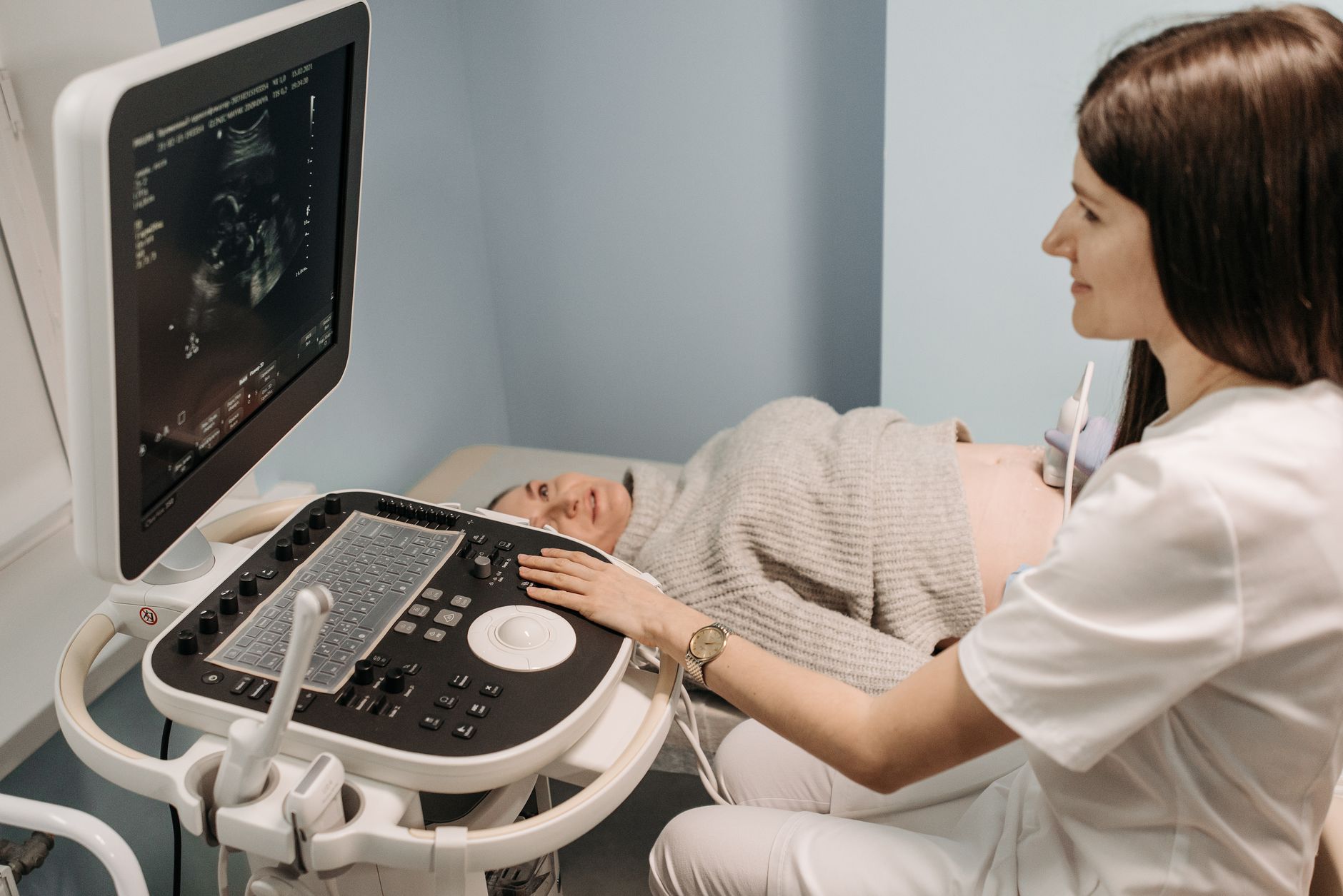
Typical ultrasound view when the sonographer records the amniotic fluid index.
How often should amniotic fluid be measured during pregnancy?
For most low‑risk pregnancies, amniotic fluid is assessed at three key points:
Mid‑pregnancy anatomy scan (18‑22 weeks): This comprehensive ultrasound includes an AFI or DVP check as part of the standard anatomy survey.
Late‑second‑trimester follow‑up (28‑30 weeks): Many providers repeat the fluid assessment at the beginning of the third trimester, especially if the earlier scan showed borderline values.
Third‑trimester monitoring (34‑36 weeks): A final check before labor helps identify any late‑onset changes that could affect delivery planning.
If any of those scans reveal a value outside the normal range, the frequency typically escalates to every 1–2 weeks until the issue resolves or a delivery plan is made.
High‑risk pregnancies—such as those involving maternal hypertension, pre‑eclampsia, diabetes, fetal growth restriction, or multiple gestations—often receive more intensive monitoring. ACOG recommends that women with these risk factors have amniotic fluid evaluated at every prenatal visit after 28 weeks, which may mean weekly scans for the last six weeks of pregnancy (ACOG Practice Bulletin No. 225, 2022).
In practice, the schedule looks like this:
Low‑risk: One scan per trimester (≈ 3 total), with an extra scan if symptoms or ultrasound findings raise concern.
Moderate‑risk (e.g., mild hypertension, well‑controlled diabetes): Every 4 weeks until 28 weeks, then every 2 weeks until delivery.
High‑risk (e.g., severe pre‑eclampsia, fetal growth restriction, twins): Every 1–2 weeks after 28 weeks, sometimes even weekly.
It’s worth noting that the “frequency” is a guideline, not a hard rule. Your provider may adjust the schedule based on how your baby is growing, how you feel, and any emerging symptoms. For example, a sudden drop in fetal movements often prompts an immediate repeat ultrasound, regardless of the routine timetable.
Recent data from the NHS suggest that women who receive more frequent fluid monitoring in high‑risk pregnancies have a modest reduction in emergency cesarean rates, likely because early detection allows for planned delivery when needed.
Staying well‑hydrated can help maintain healthy amniotic fluid levels, especially if you’re at risk for oligohydramnios.
Methods of assessment: ultrasound vs. amniocentesis
Ultrasound is the first‑line, non‑invasive tool for measuring amniotic fluid. A transabdominal probe sends high‑frequency sound waves into the uterus, creating an image that lets the sonographer trace fluid pockets and calculate AFI or DVP. The technique is safe, painless, and can be repeated as often as needed without radiation exposure (NHS, 2023).
Amniocentesis, by contrast, is an invasive procedure in which a thin needle is inserted through the abdomen to withdraw a small sample of fluid. While amniocentesis can provide precise fluid volume data, its primary purpose is genetic testing (e.g., for chromosomal abnormalities) rather than routine fluid monitoring. The procedure carries a small risk of miscarriage—approximately 0.1‑0.3 % according to the CDC—so it is reserved for situations where the diagnostic benefit outweighs that risk (CDC, 2022).
In most scenarios, clinicians rely on serial ultrasounds to track trends. Only when ultrasound findings are ambiguous, when there is a suspicion of infection, or when a concurrent genetic indication exists will an amniocentesis be considered. Even then, the fluid volume measured during amniocentesis is used as a confirmatory data point rather than a primary screening tool.
Emerging point‑of‑care ultrasound devices are being studied for use in low‑resource settings, but current guidelines from ACOG still recommend a certified sonographer for accurate AFI/DVP measurement.
When low fluid (oligohydramnios) calls for action: triggers and interventions
Oligohydramnios is defined by an AFI < 5 cm or a DVP < 2 cm. The decision to intervene hinges on three main factors:
Gestational age: Earlier gestations (before 28 weeks) are more vulnerable because the fetus relies on fluid for lung development.
Fetal growth: If the baby is already small for gestational age, low fluid may exacerbate growth restriction.
Maternal symptoms: Persistent contractions, decreased fetal movements, or signs of dehydration raise concern.
When any of those red flags appear, clinicians usually move from observation to active management. Common interventions include:
1. Maternal hydration
Increasing oral fluid intake (2–3 L of water per day) is the simplest first step. Studies published by the American College of Obstetricians and Gynecologists (ACOG 2022) show that short‑term maternal hydration can raise the DVP by up to 1 cm in many cases, especially when dehydration is the primary cause.
2. Maternal positioning
Lying on the left side improves uterine blood flow, which can boost fetal urine production and, consequently, amniotic fluid volume. Some providers advise a “left‑lateral rest” for several hours a day if oligohydramnios is detected (NHS, 2023).
3. Amnioinfusion
In more severe cases—particularly when low fluid is linked to labor complications—an amnioinfusion may be performed. This involves gently introducing sterile saline into the amniotic sac during labor, typically via a transcervical catheter. The procedure can reduce cord compression and improve the chance of a vaginal delivery, but it is reserved for hospitals with experienced obstetric teams (SMFM, 2023).
4. Treating underlying maternal conditions
If hypertension, diabetes, or medication use (e.g., ACE inhibitors) is contributing to low fluid, adjusting those treatments can help normalize levels. Your obstetrician will coordinate with your primary care provider or endocrinologist to find the safest regimen.
5. Close surveillance
When interventions stabilize fluid volume, the care plan often shifts to weekly or bi‑weekly ultrasounds, along with daily fetal movement counting, until delivery. In some cases, early delivery (induction or cesarean) may be recommended if the fluid does not improve and fetal distress emerges.
Overall, the goal is to correct the fluid deficit while avoiding unnecessary preterm birth. Most families experience a gradual improvement with conservative measures, and only a minority require invasive procedures.
Recent ACOG guidance emphasizes that any decision to deliver early should be based on a combination of fluid trends, fetal growth curves, and maternal well‑being, rather than a single AFI value alone.
Managing too much fluid (polyhydramnios): risks and options
Polyhydramnios—AFI > 25 cm or DVP > 8 cm—can also pose challenges. Excess fluid stretches the uterus, increasing the risk of preterm labor, placental abruption, and, in severe cases, maternal respiratory discomfort.
Common causes include maternal diabetes, fetal gastrointestinal obstruction, or twin‑to‑twin transfusion syndrome (TTTS) in multiples. Management strategies are tailored to the underlying cause and gestational age:
Optimizing maternal glucose: Tight glycemic control in diabetic pregnancies often reduces excess fluid (American Diabetes Association, 2023).
Therapeutic amnioreduction: A controlled removal of fluid via needle aspiration can relieve uterine over‑distension and lower the risk of preterm labor. This is typically done after 28 weeks and is most effective when the fluid excess is severe (SMFM, 2023).
Medication: In select cases, indomethacin (a prostaglandin inhibitor) is used to reduce fetal urine output, but its use is limited to before 32 weeks because of potential fetal kidney effects (FDA, 2022).
Close monitoring: Frequent ultrasounds (often weekly) and maternal symptom checks are essential to catch rapid changes.
Most cases of mild polyhydramnios resolve on their own, especially when the underlying cause is well‑controlled. Nonetheless, your provider will keep a close eye on the trend, because rapid fluid shifts can signal emerging complications.
Guidelines from the Royal College of Obstetricians and Gynaecologists (RCOG 2021) note that maternal discomfort or respiratory compromise should prompt earlier intervention, even if the AFI is only modestly elevated.
Special situations: twins, dehydration, steroids, and other considerations
Multiple gestations naturally produce more amniotic fluid, yet they also carry a higher risk of both oligohydramnios and polyhydramnios. For twins, providers usually assess each sac separately, reporting an AFI for each fetus or a combined measurement. The typical monitoring schedule for twins is:
First anatomy scan (18‑22 weeks) with individual AFI/DVP for each fetus.
Follow‑up scans at 28 weeks and 34 weeks, then every 2 weeks until delivery.
Because twins often share a common placenta, fluid imbalances can affect both babies simultaneously, making vigilant surveillance crucial.
Dehydration—whether from excessive caffeine, hot weather, or vomiting—can temporarily lower amniotic fluid. A simple trial of increased water intake, coupled with a short‑term repeat ultrasound (often within 24–48 hours), can confirm whether hydration alone corrects the low reading.
When steroids are administered for fetal lung maturity (typically between 24 and 34 weeks), they can cause a transient dip in fluid volume. Guidelines from the Royal College of Obstetricians and Gynaecologists (RCOG 2021) advise a repeat ultrasound 48 hours after the steroid course to reassess the AFI. Most women return to baseline levels without intervention.
Other rare triggers include prolonged rupture of membranes (PROM) and certain infections that affect fetal urine output. In each case, the underlying cause dictates the treatment plan, not the fluid number alone.
Twins often require more frequent amniotic fluid checks to ensure both babies are thriving.
How lifestyle factors influence amniotic fluid volume
Beyond medical conditions, everyday habits can sway fluid levels. Adequate daily water intake—typically 2.5–3 L for pregnant people—supports optimal amniotic fluid production. A BMJ systematic review (2021) found that women who increased fluid consumption by at least 500 mL per day showed a modest but statistically significant rise in DVP measurements within a week.
Conversely, high caffeine intake (more than 300 mg per day, roughly three cups of coffee) may increase urinary loss and modestly lower amniotic fluid, especially when combined with poor overall hydration. The NHS advises limiting caffeine to 200 mg per day during pregnancy. Reducing caffeine and drinking extra water can help keep fluid levels stable.
Nutrition also plays a role. A diet rich in fruits, vegetables, and lean protein supplies the electrolytes needed for proper fluid balance. Some clinicians suggest a modest increase in potassium‑rich foods (bananas, sweet potatoes) to support renal function, though evidence is still emerging.
Regular, moderate exercise—such as a 30‑minute walk—has not been shown to adversely affect fluid volume and can improve overall placental blood flow. In fact, a recent ACOG statement notes that gentle activity may help maintain healthy uterine perfusion, indirectly supporting amniotic fluid production.
What to expect after interventions: monitoring and delivery planning
Once an intervention is initiated—whether it’s hydration, positional therapy, or a procedural amnioreduction—your provider will schedule follow‑up ultrasounds to track the trend. Typically, a repeat scan is performed within 48–72 hours for hydration or positioning measures, and within one week after more invasive procedures.
If fluid levels improve and remain stable, the care plan may revert to routine trimester scans. However, if the abnormality persists, discussions about timing of delivery become central. For oligohydramnios after 34 weeks, many obstetricians consider induction to avoid fetal distress, while for polyhydramnios with severe uterine over‑distension, a planned cesarean may be recommended to reduce the risk of precipitous labor.
Throughout this period, you’ll likely be asked to keep a daily fetal movement log, monitor your own weight gain, and report any new symptoms such as sudden swelling, shortness of breath, or abdominal pain. These self‑monitoring steps help your team catch complications early, without relying solely on imaging.
When a delivery plan is being shaped, providers often use the fluid trend alongside other indicators—like biophysical profile scores and Doppler flow studies—to decide whether a scheduled induction or a watch‑and‑wait approach is safest.
Interpreting serial measurements: trends vs. single values
A single AFI or DVP reading provides a snapshot, but clinicians place far more weight on how the numbers change over time. A gradual decline of a few centimeters over several weeks may be reassuring if the baby continues to grow appropriately, whereas a rapid drop of more than 2 cm within a few days can signal impending distress.
Because of this, many providers chart fluid measurements alongside fetal growth curves on the same ultrasound report. This visual pairing helps both the care team and the family see the broader picture. If you notice that your fluid values are trending downward, bring the chart to your next appointment and ask how the trend fits with your overall pregnancy plan.
Medication effects on amniotic fluid
Certain prescription medications can influence amniotic fluid volume. For example, ACE inhibitors and angiotensin‑II receptor blockers are known to reduce fetal kidney function, leading to oligohydramnios; they are therefore contraindicated in pregnancy (FDA, 2022). Conversely, some antidiabetic drugs, such as insulin, help control maternal glucose and can indirectly reduce polyhydramnios caused by diabetic hyperglycemia.
If you are taking any chronic medication, discuss its safety with your obstetrician early in pregnancy. Your provider may switch you to a pregnancy‑compatible alternative or monitor your amniotic fluid more closely if a drug cannot be changed.
Doctor's note
From our medical team: Amniotic fluid measurements are a valuable, non‑invasive window into your baby's health. If your scan shows a borderline value, most clinicians will first suggest simple lifestyle tweaks—extra water, left‑side rest, and a short‑term repeat scan—before moving to more invasive options. Always discuss any new symptoms (like reduced fetal movement or painful contractions) with your provider promptly, and remember that a single measurement is only a snapshot; trends over time are what truly guide care.
🔢 Ready to crunch your numbers? Use our AFI / SDP Interpreter for a personalized result in seconds.
Myth vs. fact
Myth: “If my AFI is slightly low, I should schedule an emergency C‑section right away.”
Fact: A mildly low AFI (e.g., 4.5 cm) often improves with hydration and positional therapy. Immediate delivery is reserved for cases where low fluid is accompanied by fetal distress or other high‑risk factors.
Myth: “Amniotic fluid can be measured with a regular home ultrasound device.”
Fact: Accurate AFI/DVP measurement requires a certified sonographer and a high‑resolution obstetric ultrasound. Home devices cannot reliably capture the necessary detail.
Myth: “Polyhydramnios is always dangerous and requires medication.”
Fact: Mild polyhydramnios is common and often resolves with proper maternal glucose control. Medications are only used in severe or rapidly progressing cases.
Key takeaways
Amniotic fluid monitoring is routine; most low‑risk pregnancies have an ultrasound each trimester.
Normal AFI ranges from 5 – 25 cm; normal DVP ranges from 2 – 8 cm across all trimesters.
Intervention is usually triggered when AFI < 5 cm or DVP < 2 cm (low) or AFI > 25 cm or DVP > 8 cm (high).
First‑line treatments for low fluid include increased hydration, left‑side rest, and close repeat scanning.
High‑risk pregnancies (e.g., hypertension, diabetes, twins) often receive fluid checks every 1–2 weeks after 28 weeks.
Always call your provider if you notice reduced fetal movement, painful contractions, or a sudden change in fluid‑related symptoms.
Frequently asked questions
What is the normal amniotic fluid index (AFI) range?
The standard range is 5 – 25 cm throughout pregnancy; values below 5 cm suggest oligohydramnios, while values above 25 cm indicate polyhydramnios.
How often should my doctor check amniotic fluid during pregnancy?
For low‑risk pregnancies, expect an ultrasound at the anatomy scan (18‑22 weeks), a late‑second‑trimester scan (≈ 28 weeks), and a third‑trimester scan (≈ 34‑36 weeks). High‑risk situations may require weekly or bi‑weekly checks after 28 weeks.
What triggers an intervention for low amniotic fluid?
Intervention is usually considered when AFI < 5 cm or DVP < 2 cm, especially if the fetus shows slowed growth, if you’re past 28 weeks, or if you have symptoms like reduced fetal movement.
Can high amniotic fluid levels be dangerous?
Yes, an AFI > 25 cm can increase the risk of preterm labor, uterine over‑distension, and, in severe cases, placental abruption. Management depends on the cause and gestational age.
Is amniotic fluid monitoring done with a regular ultrasound?
Yes—most providers assess fluid during a standard obstetric ultrasound, calculating either the AFI or the deepest vertical pocket. Amniocentesis is reserved for specific diagnostic purposes.
What treatments are available if oligohydramnios is diagnosed?
First‑line measures include increased water intake, left‑side rest, and treating any underlying maternal condition. If fluid does not improve, options such as amnioinfusion or early delivery may be discussed.
Can I track amniotic fluid at home with a wearable or app?
Currently, no home device can reliably measure AFI or DVP. While some wearables claim to estimate fetal hydration, the gold standard remains a professional ultrasound performed by a certified sonographer. If you’re concerned about fluid levels, schedule a scan rather than relying on a consumer gadget.
Does caffeine affect amniotic fluid volume?
High caffeine intake (more than 300 mg per day) may increase urinary output and modestly lower amniotic fluid, especially when overall hydration is low. The NHS recommends limiting caffeine to 200 mg per day during pregnancy. Reducing caffeine and drinking extra water can help keep fluid levels stable.
What is the difference between AFI and SDP?
AFI adds fluid depths from four uterine quadrants, while SDP (single deepest pocket) records only the deepest vertical pocket. Both are accepted methods; some clinicians prefer SDP because it reduces false‑positive oligohydramnios diagnoses.
Can dehydration cause a false low amniotic fluid reading?
Yes—temporary dehydration can lower the DVP on an ultrasound. A short‑term hydration trial followed by a repeat scan within 24–48 hours can distinguish true oligohydramnios from a dehydration artifact.
When to call your doctor
If you notice any of the following, contact your obstetric provider right away: sudden decrease in fetal movements, persistent abdominal pain or contractions, vaginal bleeding, leakage of fluid, fever, or a rapid weight gain with swelling. This article is for informational purposes only and does not replace personalized medical advice.
References
American College of Obstetricians and Gynecologists. “Practice Bulletin No. 225: Ultrasonography in Pregnancy.” 2022.
National Health Service (NHS). “Amniotic fluid – normal range and tests.” Updated 2023.
Royal College of Obstetricians and Gynaecologists. “Guidelines on the use of antenatal steroids and amniotic fluid monitoring.” 2021.
Centers for Disease Control and Prevention (CDC). “Maternal and Infant Health: Amniotic Fluid Abnormalities.” 2022.
World Health Organization (WHO). “Maternal health: recommendations for prenatal care.” 2022.
Society for Maternal‑Fetal Medicine (SMFM). “Management of oligohydramnios and polyhydramnios.” 2023.
British Medical Journal (BMJ). “Maternal hydration and amniotic fluid volume: a systematic review.” 2021.
American Diabetes Association. “Standards of Care in Diabetes—2023.”
Food and Drug Administration (FDA). “Indomethacin Use in Pregnancy – Safety Information.” 2022.
National Institute for Health and Care Excellence (NICE). “Hydration and caffeine guidelines for pregnancy.” 2023.
American College of Obstetricians and Gynecologists. “Guidance on Maternal Hydration and Fetal Well‑Being.” 2022.
Society for Maternal‑Fetal Medicine. “Medication Safety in Pregnancy.” 2023.
Editor's pick for this topic
About the Author
When Shubhra Mishra was expecting her first child in 2016, she was overwhelmed by conflicting food advice — one site said yes, another said never. By the time her second baby arrived in 2019, she realized millions of mothers face the same confusion.
That sparked a five-year journey through clinical nutrition papers, cultural diets, and expert conversations — all leading to BumpBites: a calm, compassionate space where science meets everyday motherhood.
Her long-term vision is to build a global community ensuring safe, supported, and free deliveriesfor every mother — because no woman should face pregnancy alone or uninformed. 🌿
🌍 Stand with mothers, shape safer guidance
Join a small circle of experts who review BumpBites articles so expecting parents everywhere can decide with confidence.