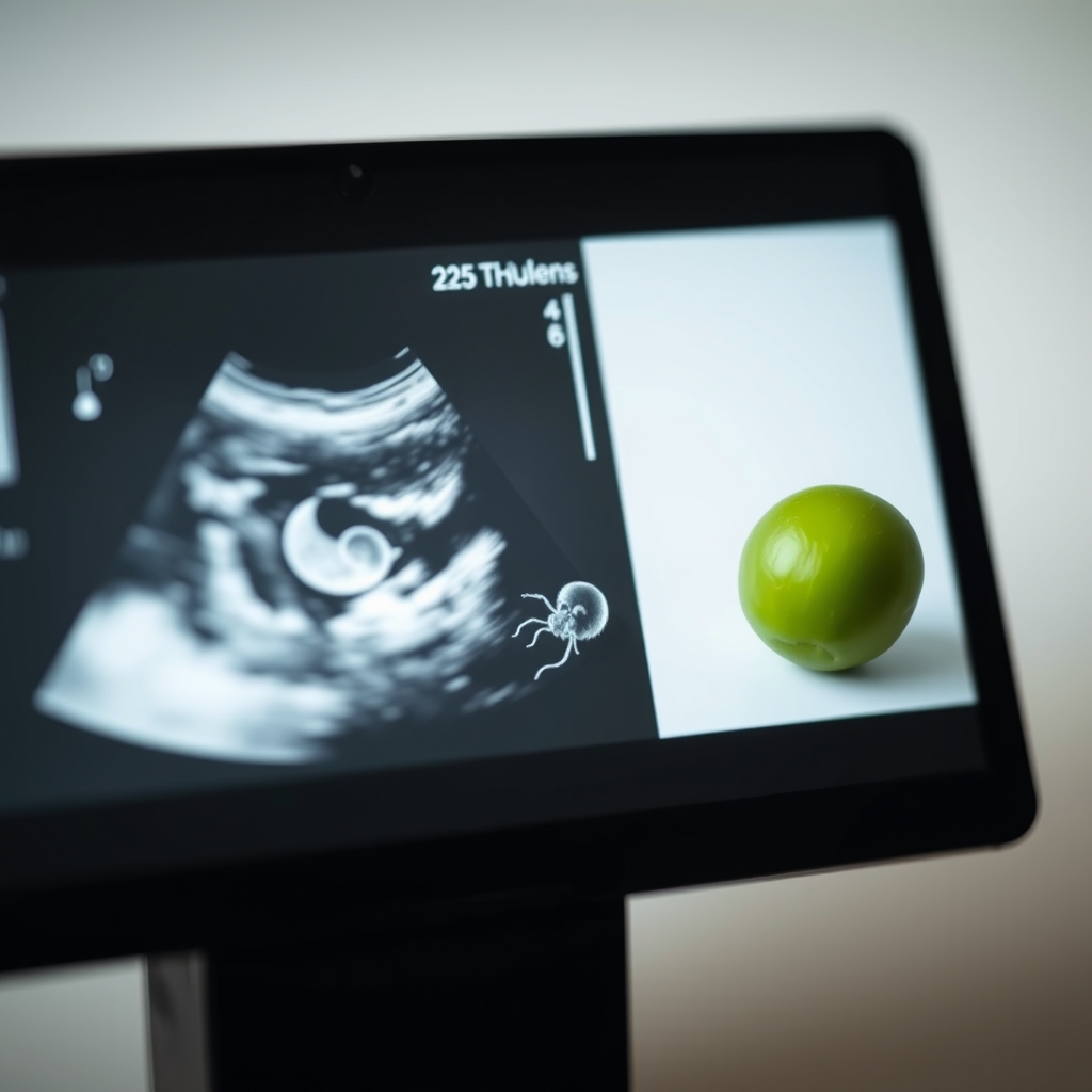
At 7 weeks pregnant, baby size is about the size of a blueberry, measuring around 10mm in length, with major organs starting to develop
By Shubhra Mishra — a mom of two who turned her own confusion during pregnancy into BumpBites, a global mission to make food choices clear, safe, and stress-free for every expecting mother. 💛
Check whether any food is safe during pregnancy with the BumpBites Food Safety Checker.
Quick take: At 7 weeks pregnant, the embryo measures about 0.5 inches (12–13 mm) long, weighs roughly 0.04 grams, and looks like a tiny bean‑shaped structure with a developing heartbeat. Most mothers can’t feel movement yet, but a faint heartbeat can often be heard on a transvaginal ultrasound. Expect mild nausea, breast tenderness, and a sense that something is “growing.”
It’s 2 a.m., you’re curled up on the couch, and a sudden wave of nausea makes you wonder: “How big is my baby right now?” You’ve just hit the 7‑week mark and the flood of questions feels overwhelming. The good news is that by week 7, there’s a clear picture of how big your embryo is, what it’s doing, and what you might be feeling.
In this guide we’ll give you the exact measurements in inches, centimeters, and even a pea‑size comparison. We’ll walk through what the ultrasound looks like, the tiny heartbeat that may be audible, the milestones your baby is hitting, and the common symptoms you can expect. We’ll also compare week 7 to week 6 and week 8, show you how to track growth, and answer the most‑asked questions so you can feel confident in this early stage of pregnancy.
All the information is drawn from the latest guidance from ACOG, the NHS, and the CDC, and we’ll point out where you might want to ask your own provider for clarification.
What is the size of a 7‑week pregnant baby in inches, centimeters, and pea‑size comparison?
At the start of week 7 (which is actually 6 weeks after fertilization), the embryo measures roughly 0.5 inches long, which translates to 12–13 mm or about 0.5 cm. In practical terms, it’s about the size of a small pea or a lentil. This tiny length is measured from crown (top of the head) to tail (bottom of the body) and is called the crown‑rump length (CRL).
Because many expectant parents think in both metric and imperial units, we’ve created a quick size chart:
Measurement
Inches
Centimeters
Common Comparison
Crown‑rump length
≈ 0.5 in
≈ 12–13 mm
Small pea or lentil
Head diameter
≈ 0.2 in
≈ 5 mm
Grain of rice
These numbers are averages; individual embryos can be a little larger or smaller. The size chart is useful when you’re looking at an ultrasound image or simply trying to picture the growth.
Imagine a tiny pea—your baby is about that size at 7 weeks.
How many weeks old is a 7‑week fetus compared to a newborn?
The term “7‑week fetus” can be confusing because the word “fetus” technically applies after the end of the 8th week of gestation. Nonetheless, many people use “fetus” and “embryo” interchangeably in casual conversation. At 7 weeks gestational age, the embryo is roughly the same age as a newborn who is just a few days old in terms of cell division, but the developmental stage is far earlier.
In other words, a 7‑week embryo has been growing for about 5 weeks since conception. By comparison, a full‑term newborn has completed about 38 weeks of gestation. The gap is enormous: the newborn has fully formed organs, lungs, and a brain capable of basic reflexes, while the 7‑week embryo is still establishing its basic body plan.
If you picture a newborn’s tiny hand, that’s the level of development you’ll see in the next few weeks—not now. The key takeaway is that “7 weeks” refers to the gestational age counted from the first day of the last menstrual period, not the actual age of the embryo.
What does a 7‑week embryo look like on an ultrasound, and how can I use it as a size reference?
By week 7, most providers perform a transvaginal ultrasound to confirm the pregnancy and check the heartbeat. On the screen, the embryo appears as a tiny, bean‑shaped structure with a clear gestational sac surrounding it. You’ll see a small dark spot that represents the yolk sac, which supplies nutrients early on.
The embryo’s outline is usually visible as a faint gray silhouette, and you may be able to spot the beginning of the neural tube (the future brain and spinal cord) as a thin line. The most exciting part for many parents is spotting a flickering red dot—this is the heartbeat, typically beating between 90–110 bpm at this stage.
Because the ultrasound image is calibrated, you can use the on‑screen ruler to compare the embryo’s length directly to the 0.5‑inch measurement from the size chart. Many clinics provide a printed image with a scale, allowing you to hold it next to a pea or a small coin for a tangible reference.
Below is a typical ultrasound image reference guide:
Week
CRL (mm)
Visual Cue
6
5–6
Pinhead
7
12–13
Pea‑size bean
8
16–20
Small blueberry
How much does a 7‑week pregnant baby weigh, and what is the average weight in grams?
The embryo’s weight at 7 weeks is minuscule—about 0.04 grams, which is roughly the weight of a single grain of rice. In metric terms, that’s 40 milligrams. Because the weight is so low, most scales cannot measure it, and the number is based on scientific estimates rather than direct weighing.
Even though the weight is tiny, the rapid cell proliferation means the embryo is doubling in size every few days. The increase in mass is primarily due to the formation of new tissue and the early development of the circulatory system, not because of fat accumulation.
For perspective, a full‑term baby typically weighs between 2.5 kg and 4 kg. So the 7‑week embryo’s weight is less than one‑hundredth of a percent of the final birth weight. This helps put into context why you can’t “feel” the baby yet; the mass is simply too small.
What are the developmental milestones at 7 weeks gestation, including brain changes?
Week 7 is a busy time for organogenesis—the period when the embryo’s major organs begin to form. Here are the key milestones:
Neural tube closure: The tube that will become the brain and spinal cord has sealed, reducing the risk of neural tube defects.
Brain development: The forebrain (prosencephalon) starts to differentiate, laying the groundwork for future cerebral hemispheres. Early neurons are beginning to migrate.
Heart: The heart has begun to beat regularly (90–110 bpm) and is beginning to loop, a critical step in forming chambers.
Eyes and ears: Optic vesicles (precursors to eyes) appear, and the inner ear begins to develop, which will later enable balance.
Limb buds: Small protrusions that will become arms and legs are visible, though fingers and toes are not yet formed.
Yolk sac: Provides early nutrition and is clearly visible on ultrasound.
These milestones are confirmed by ACOG’s guidelines for early prenatal care and are the basis for most 7‑week ultrasound assessments.
Can I feel my 7‑week pregnant baby move yet, and what should I expect?
Most people cannot feel fetal movement at 7 weeks. The first sensations—often described as “quickening”—typically occur between 16 and 25 weeks, depending on the mother’s body type and whether it’s a first pregnancy.
That said, some women report a vague flutter or a “butterfly” feeling as early as 12 weeks. At 7 weeks, the embryo’s muscles are just beginning to form, and there isn’t enough mass to generate a noticeable movement. If you do feel a sensation, it’s more likely to be gas or a muscle twitch, not the baby.
When you do start to feel movement later in the second trimester, it’s a reassuring sign that the baby’s neuromuscular system is functioning. Until then, focus on other health markers such as heartbeat detection and growth measurements.
What are the common symptoms for a woman at 7 weeks pregnant?
Week 7 is often marked by the “morning sickness” phase, though symptoms can occur at any time of day. Typical symptoms include:
Nausea and vomiting: Hormonal changes (especially hCG) trigger the “morning sickness” wave.
Breast tenderness: Increased blood flow and hormonal shifts make the breasts feel sore, swollen, or tingly.
Fatigue: The body is working hard to support the rapidly developing embryo.
Frequent urination: The growing uterus puts pressure on the bladder.
Food cravings or aversions: Hormonal fluctuations can alter taste preferences.
Mood swings: Emotional ups and downs are common as the body adapts.
While most of these symptoms are normal, any severe vomiting, intense abdominal pain, or spotting should prompt a call to your provider (see “When to call your doctor” below).
How does a 7‑week embryo compare to a 6‑week and an 8‑week embryo?
Understanding the progression from week 6 to week 8 helps you visualize how quickly things change.
Small blueberry size, facial features start to form
At week 6, the embryo is still a tiny disc; by week 7, the heart beats and limb buds emerge; by week 8, the head becomes more distinct, facial prominences appear, and the embryo resembles a tiny bean with a recognizable “head.” This rapid growth underscores why early prenatal visits are crucial for monitoring development.
When does the baby's heartbeat start at 7 weeks, and how is it detected?
The fetal heartbeat usually becomes detectable between 6 and 7 weeks gestation using a transvaginal ultrasound. A Doppler fetal monitor (the handheld “heartbeat” device) may not pick up the sound until around 10–12 weeks, but the ultrasound can see the heartbeat as early as 6 weeks.
During the ultrasound, the technician will look for a flickering red dot that corresponds to the heart’s rhythmic motion. The normal range at 7 weeks is 90–110 bpm. If the heartbeat is absent or irregular, the provider may schedule a follow‑up scan to reassess.
Detecting the heartbeat early provides reassurance that the embryo is developing properly and helps establish a baseline for later monitoring.
Early heartbeat detection is a key milestone at 7 weeks.
How to track baby growth at 7 weeks pregnant and what to expect in prenatal appointments
Tracking growth at this early stage relies on a combination of ultrasound measurements, hormone level checks, and symptom monitoring.
Ultrasound: Your provider will measure the crown‑rump length (CRL) and compare it to the standard growth chart. Consistency with the chart indicates normal development.
hCG levels: Blood tests for human chorionic gonadotropin can help confirm that the pregnancy is progressing. Levels typically double every 48–72 hours in early weeks.
Symptom diary: Keeping a simple log of nausea, breast tenderness, and energy levels can help you recognize patterns and discuss any concerns with your provider.
First prenatal visit: Usually scheduled between weeks 8–10, this appointment includes a full physical exam, blood work, and a detailed ultrasound to confirm the embryo’s age and heartbeat.
When you attend your 7‑week check‑up, expect the clinician to ask about any bleeding, severe pain, or unusual symptoms. They’ll also discuss nutrition, prenatal vitamins (especially folic acid), and lifestyle recommendations to support early development.
From our medical team: “At seven weeks, the embryo’s heartbeat is a reassuring sign of viability, but remember that tiny variations in size are normal. If your ultrasound shows a CRL within the expected range, you’re on track. Keep taking your prenatal vitamin, stay hydrated, and reach out to your provider if you notice heavy bleeding or persistent severe nausea.”
Myth vs. fact
Myth: “If I can’t feel my baby move at 7 weeks, something is wrong.”
Fact: Movement isn’t felt until the second trimester. At 7 weeks the embryo is too small and its muscles are just forming.
Myth: “All babies are the same size at 7 weeks.”
Fact: While the average CRL is about 0.5 inches, natural variation of a few millimeters is completely normal.
Myth: “A heartbeat must be heard with a Doppler device at 7 weeks.”
Fact: The heartbeat is usually visualized on a transvaginal ultrasound; a Doppler may not pick it up until later weeks.
Key takeaways
The 7‑week embryo is about 0.5 inches (12–13 mm) long—roughly the size of a pea.
Weight is roughly 0.04 grams (40 mg), far too tiny to feel.
A heartbeat of 90–110 bpm can often be seen on a transvaginal ultrasound.
Key milestones include neural tube closure, early brain formation, and limb‑bud emergence.
Common symptoms are nausea, breast tenderness, and frequent urination; severe pain or bleeding warrants a call to your provider.
Movement isn’t felt until later (≈ 16–25 weeks), so lack of sensation at 7 weeks is normal.
Track growth with ultrasound CRL measurements, hCG levels, and symptom logs, and attend your first prenatal visit by weeks 8–10.
Frequently asked questions
How big is a baby at 7 weeks pregnant?
At 7 weeks, the embryo measures about 0.5 inches (12–13 mm) long, comparable to a small pea.
What does a 7‑week embryo look like on an ultrasound?
It appears as a tiny bean‑shaped silhouette within a gestational sac, often with a visible heartbeat and a small yolk sac attached.
Can I feel my baby move at 7 weeks?
No, fetal movement isn’t usually felt until the second trimester (around 16‑25 weeks); at 7 weeks the embryo is too small.
What are the signs of a healthy 7‑week pregnancy?
Key signs include a detectable heartbeat on ultrasound, steady hCG rise, mild nausea, breast tenderness, and no heavy bleeding or severe abdominal pain.
How much does a 7‑week fetus weigh?
The embryo weighs roughly 0.04 grams (about 40 mg), which is similar to the weight of a single grain of rice.
When does the baby's heartbeat become detectable?
Using a transvaginal ultrasound, the heartbeat can often be seen as early as 6 weeks, and is reliably visible by 7 weeks with a rate of 90–110 bpm.
What should I expect during a 7‑week ultrasound?
You’ll likely see the gestational sac, yolk sac, and a tiny bean‑shaped embryo with a faint heartbeat. The sonographer will measure the crown‑rump length to confirm dating.
How does a 7‑week embryo compare to a 6‑week embryo?
At week 6 the embryo is about 0.2 inches long and may not yet have a visible heartbeat. By week 7 it doubles in length, the heart beats, and limb buds become evident.
When to call your doctor
If you experience any of the following, contact your provider right away: heavy vaginal bleeding, severe abdominal pain, fever over 100.4 °F (38 °C), sudden swelling of the face or hands, or any signs of infection. This article is for general information only and does not replace personalized medical advice.
References
American College of Obstetricians and Gynecologists (ACOG). “Early Pregnancy.” Practice Bulletin No. 171, 2022.
National Health Service (NHS). “Pregnancy week by week.” Updated 2023.
Centers for Disease Control and Prevention (CDC). “Prenatal Care.” 2023.
World Health Organization (WHO). “Fetal development: first trimester.” 2021.
Mayo Clinic. “First trimester ultrasound: What to expect.” 2022.
Royal College of Obstetricians and Gynaecologists (RCOG). “Ultrasound in early pregnancy.” 2022.
American Academy of Pediatrics (AAP). “Developmental milestones: First trimester.” 2023.
Not sure about the label on 7 Weeks Pregnant Baby Size products?
Snap the ingredients list and SafeFilter checks every ingredient for your stage — only 3 free scans this month, then you're locked until reset. Unlimited from $7/mo or $50/yr.
When Shubhra Mishra was expecting her first child in 2016, she was overwhelmed by conflicting food advice — one site said yes, another said never. By the time her second baby arrived in 2019, she realized millions of mothers face the same confusion.
That sparked a five-year journey through clinical nutrition papers, cultural diets, and expert conversations — all leading to BumpBites: a calm, compassionate space where science meets everyday motherhood.
Her long-term vision is to build a global community ensuring safe, supported, and free deliveriesfor every mother — because no woman should face pregnancy alone or uninformed. 🌿
🌍 Stand with mothers, shape safer guidance
Join a small circle of experts who review BumpBites articles so expecting parents everywhere can decide with confidence.