At 6 weeks pregnant, your baby is about the size of a sweet pea (0.25 inches). Learn about fetal development, symptoms, and what to expect this week.
By Shubhra Mishra — a mom of two who turned her own confusion during pregnancy into BumpBites, a global mission to make food choices clear, safe, and stress-free for every expecting mother. 💛
Check whether any food is safe during pregnancy with the BumpBites Food Safety Checker.
Quick take: At six weeks pregnant, the embryo is about 5 mm long (≈0.2 in) and weighs roughly 0.05 g—about the size of a lentil or a raspberry. Most embryos fall within a narrow size range, and any major deviation should be discussed with your provider.
It’s 2 a.m., you’re curled up with a cup of tea, and a vague “baby size” worry is keeping you up. You’ve just read that you’re six weeks along and wonder whether the tiny figure you saw on the ultrasound is “normal.” You’re not alone—most expectant parents ask the same question. Below, we break down exactly how big a six‑week baby is, what it looks like, how it’s measured, and what milestones to expect. By the end you’ll have a clear picture, a handy size chart, and a list of red‑flag symptoms that warrant a call to your provider.
We’ll walk through the exact length in millimeters, compare it to familiar fruits, describe the ultrasound image, and explain the embryo’s development at this early stage. We’ll also cover normal size ranges, weight estimates, and how the six‑week size differs from the five‑week stage. All information is drawn from reputable bodies such as the American College of Obstetricians and Gynecologists (ACOG), the UK’s National Health Service (NHS), and the World Health Organization (WHO). Remember, this guide is informational only—always discuss personal concerns with your healthcare team.
How big is a baby at 6 weeks pregnant?
At six weeks gestation, what most people call a “baby” is technically an embryo—a cluster of delicate cells beginning to form the foundations of the future body. The average embryo measures about 5 mm in length, which is roughly the size of a grain of rice or a tiny raspberry. In practical terms, it fits comfortably on the tip of a standard adult finger.
Why the “average” matters: ultrasound studies compiled by ACOG indicate that 95 % of embryos measured between 4 mm and 6 mm at six weeks. The slight variation comes from factors such as exact dating (based on the first day of your last menstrual period), maternal body type, and the precision of the ultrasound equipment.
Most expectant parents discover the size during a transvaginal ultrasound, usually scheduled between 5 and 7 weeks. During this scan, the sonographer measures the crown‑rump length (CRL)—the distance from the top of the head (crown) to the buttocks (rump). This single dimension is the most reliable way to estimate gestational age at this point.
Below is a quick reference chart for the six‑week embryo size:
Gestational Age
Crown‑Rump Length
Typical Appearance
5 weeks
≈ 2 mm
Small “bean‑shaped” speck
6 weeks
≈ 5 mm (0.2 in)
Visible tail and beginning of head
7 weeks
≈ 9 mm
More defined head and limb buds
Even though a 5‑mm embryo sounds minuscule, it already houses the beginnings of the brain, spinal cord, heart, and the first signs of limb formation. The next sections dive into the exact length, weight, and visual cues you can expect on your ultrasound.
Seeing that tiny speck for the first time can feel both magical and nerve‑wracking. If you’re wondering whether the size you see is “too big” or “too small,” remember that a few millimeters difference is usually just a matter of dating accuracy, not an indication of trouble.
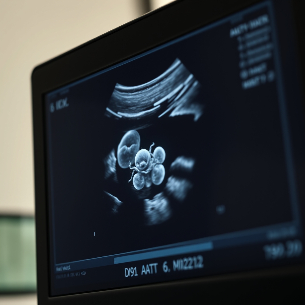
Typical six‑week ultrasound view: a tiny bright speck representing the embryo.
What is the length of a 6‑week embryo?
The crown‑rump length (CRL) at six weeks is precisely measured at about 5 mm, which translates to 0.2 inches. This measurement is taken from the top of the embryo’s head (the crown) down to the end of the torso (the rump). Because the embryo’s tail is still extending, the CRL does not include the tail itself.
How clinicians obtain this number: During a transvaginal ultrasound, the sonographer captures a thin, high‑frequency image that provides the resolution needed to see a 5‑mm structure. They then use built‑in calipers on the ultrasound machine to trace the CRL. The accuracy of this technique is high—studies in the Journal of Ultrasound in Medicine report a margin of error under 0.5 mm when performed by trained professionals.
It’s also worth noting that the embryo’s length increases by roughly 1 mm per week during the first trimester. So, if you had an ultrasound at five weeks showing a 2‑mm CRL, a one‑week later scan would typically show around 5 mm, confirming steady growth.
While the average is 5 mm, some embryos may measure 4 mm or 6 mm. A 4‑mm embryo at six weeks is still within normal limits, especially if the dating is based on the last menstrual period rather than a prior scan. Conversely, a 6‑mm measurement may suggest the pregnancy is slightly further along than initially thought, prompting a re‑evaluation of gestational dating.
In practice, the exact length guides clinicians in scheduling future appointments, ordering necessary labs, and reassuring parents that the pregnancy is progressing as expected.
Because the CRL is the most reliable single metric in early pregnancy, many providers will use it to calculate estimated due dates, rather than relying solely on calendar dates.
What are the 6‑week baby size measurements in centimeters and inches?
When you convert the 5 mm CRL to more familiar units, you get 0.5 cm (half a centimeter) and 0.2 inches. Though the numbers seem tiny, visualizing them against everyday objects helps put the size into perspective.
In the metric system, 0.5 cm is roughly the thickness of a standard pencil eraser or the width of a dime. In the imperial system, 0.2 inches matches the height of a grain of sand stacked three high. These comparisons help you gauge how much the embryo will grow in the weeks ahead—by the end of the first trimester, it will be about 2 cm (0.8 in), roughly the size of a blueberry.
Below is a concise size conversion table you can keep on your phone:
Measurement
Millimeters
Centimeters
Inches
Crown‑Rump Length
5 mm
0.5 cm
0.20 in
Estimated Weight
≈ 0.05 g
≈ 0.00005 kg
≈ 0.0018 oz
These numbers are averages; slight variations are normal. The key takeaway is that at six weeks, the embryo is still microscopic, and most expectant mothers will not feel any movement yet.
Clinicians also use these measurements to adjust medication dosages that may affect the embryo, such as folic acid supplements, ensuring the right amount is given at the right developmental stage.
6 weeks pregnant baby size compared to a fruit
One of the most popular ways clinicians help parents visualize early pregnancy is by comparing the embryo to familiar foods. At six weeks, the embryo’s size is often likened to a lentil, raspberry, or small blueberry. All three have a diameter of roughly 5 mm, making them perfect visual analogues.
Why fruit? The comparison is intuitive, non‑clinical, and instantly understandable. For instance, a fresh raspberry has a slightly irregular shape, which mirrors the embryo’s early, somewhat curved form. A lentil, on the other hand, is more uniform and can illustrate the roundness of the embryo’s head.
In practice, many ultrasound clinics display a small fruit next to the monitor when explaining the scan to patients. This tactile reference helps reduce anxiety by turning an abstract measurement into something concrete you can hold in your hand.
Here’s a quick list of fruit comparisons for each week of early pregnancy, useful for future reference:
5 weeks: a poppy seed or a tiny pea.
6 weeks: a lentil, raspberry, or small blueberry.
7 weeks: a kidney bean or a large grape.
8 weeks: a walnut or an olive.
These visual cues are especially helpful when sharing your ultrasound images with family members who may not understand medical terminology.
Different cultures may favor other analogies—some providers use a grain of sand or a small almond. The goal is always the same: to give you a mental picture you can easily recall.
What does a 6‑week baby look like on ultrasound?
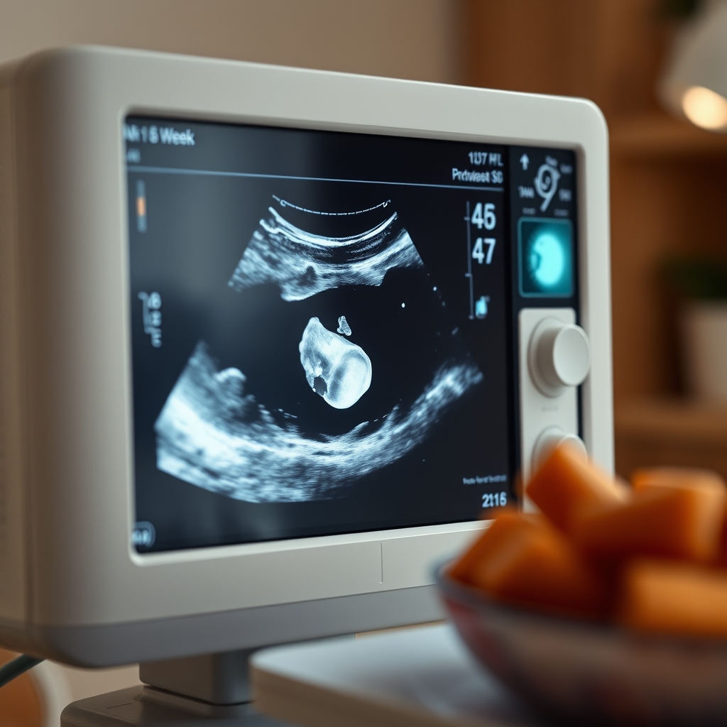
During a transvaginal ultrasound at six weeks, the image appears as a bright, tiny speck against a dark background. This speck represents the embryo’s head, and you may also see a faint curved line indicating the beginning of the tail. A faint, flickering dot—a nascent heartbeat—may be visible if the equipment is high‑resolution and the embryo is well‑positioned.
The most recognizable feature at this stage is the gestational sac, a fluid‑filled bubble that surrounds the embryo. Inside the sac, the embryo’s head is usually larger than the rest of the body, giving it a “bean‑shaped” appearance. The yolk sac, a small circular structure, appears adjacent to the embryo and supplies early nutrients.
While the image is small, it provides crucial information:
Heartbeat detection: A rhythmic flicker at 90–110 beats per minute confirms viability. The CDC notes that most embryos show a heartbeat by six weeks.
Yolk sac size: A yolk sac measuring 2–5 mm is typical; larger sizes can signal potential issues.
Location: The embryo should be centrally located within the gestational sac; an off‑center position may warrant closer monitoring.
Because the embryo is so tiny, the image can be a source of both wonder and worry. Remember that a clear view depends on several factors—maternal body habitus, bladder fullness, and the skill of the sonographer.
If you’re uncertain about what you see, ask the sonographer to point out the key structures on the screen and to explain any measurements they record. Having a printed copy of the image can also be helpful for future reference.
The six‑week embryo appears as a tiny bright spot; a faint flicker may indicate the first heartbeat.
Development milestones of a 6‑week embryo
Even though the embryo is only a few millimeters long, a cascade of critical events is already under way. By the end of the sixth week, the following structures are forming or have begun to function:
Heart tube: The primitive heart tube folds and starts beating rhythmically at about 90–110 bpm.
Neural tube: The beginning of the brain and spinal cord—known as the neural tube—closes, a process essential for proper nervous system development.
Limb buds: Small protrusions appear on either side of the embryo, the precursors to arms and legs.
Eye pits: Shallow depressions that will later become eyes.
Yolk sac: Provides early nutrition and blood cell formation.
Placental precursor: The chorionic villi begin to develop, laying the groundwork for the placenta.
According to the NHS and ACOG, the presence of a detectable heartbeat at six weeks is a reassuring sign of viability. However, the heart is still a simple tube; it will not have distinct chambers until about eight weeks.
These milestones explain why many parents feel a mix of excitement and anxiety when they first see the tiny speck on the screen. While the embryo is still far from resembling a human baby, the foundations for every major organ system are being laid.
Folic acid plays a pivotal role in neural tube closure. If you haven’t started a prenatal vitamin yet, discuss it with your provider—most guidelines recommend 400–800 µg daily during the first trimester.
Understanding these developments empowers you to ask informed questions at your next prenatal visit, such as “How is the heart forming?” or “When will we see the first limb buds on the ultrasound?”
Is 6 weeks pregnant baby size normal, and what weight can be expected?
“Normal” at six weeks means the embryo’s crown‑rump length falls between 4 mm and 6 mm, with a weight roughly between 0.03 g and 0.07 g. The estimated weight of a typical six‑week embryo is about 0.05 g—comparable to the weight of a single grain of rice.
Variation is common and usually harmless. Factors influencing size include:
Dating accuracy: If your last menstrual period (LMP) date is off by a few days, the embryo may appear larger or smaller than the calendar week suggests.
Maternal BMI: Higher body mass can affect ultrasound image clarity, sometimes making the embryo appear slightly larger.
Multiple gestations: Twins or higher‑order multiples often have slightly smaller individual embryos at the same gestational age.
If an ultrasound shows an embryo outside the 4‑6 mm range, clinicians will typically repeat the scan in a week or two to confirm growth trends. A slower‑growing embryo might still be normal, especially if the dating is uncertain. However, consistent measurements that fall far below the expected range could indicate a potential miscarriage, prompting closer monitoring.
Weight is not routinely measured at this stage, but the estimated 0.05 g is derived from standard growth curves published by the WHO. These curves are widely used in both the United States and the United Kingdom to assess early fetal development.
Overall, a six‑week embryo’s size and weight are reliable markers of healthy progression when interpreted in the context of a complete prenatal assessment.
How does the 6‑week baby size compare to the 5‑week size and what growth to expect?
Between five and six weeks, the embryo’s crown‑rump length typically triples—from about 2 mm to 5 mm. This rapid growth reflects the swift formation of the heart, neural tube, and limb buds. In practical terms, you’re looking at an increase of roughly 3 mm (0.12 in) in just one week.
Growth rates continue at an approximate 1 mm per week throughout the first trimester. By eight weeks, the embryo reaches about 9 mm (0.35 in) and begins to look more recognizable, resembling a tiny tadpole with distinct head and tail.
Because the size change is so dramatic, many parents notice a significant difference when comparing their 5‑week and 6‑week ultrasound images. The 5‑week scan often shows only a faint gestational sac, while the 6‑week scan reveals the embryo’s head, faint heartbeat, and the first hint of a tail.
Understanding this growth trajectory helps set realistic expectations: you won’t feel any fetal movement until around 16‑20 weeks, but the early size milestones are crucial for confirming that the pregnancy is on track.
A ripe raspberry—about 5 mm—mirrors the size of a six‑week embryo.
From our medical team: “Seeing a tiny speck on a six‑week ultrasound can be both awe‑inspiring and nerve‑wracking. Remember that the embryo’s size is measured with a high‑resolution probe, not by how you feel. If the CRL falls within the 4‑6 mm range and a heartbeat is present, the pregnancy is generally considered healthy. Any concerns about size should be discussed with your obstetrician, who can schedule a follow‑up scan if needed.”
What factors can affect the size of a 6‑week embryo?
While genetics set the overall growth pattern, several maternal and environmental factors can subtly influence the measured size at six weeks. The most common contributors are:
Accuracy of dating: Irregular menstrual cycles or uncertain LMP dates can shift the perceived gestational age by several days, making the embryo appear larger or smaller.
Maternal nutrition: Adequate folic acid, iron, and protein intake supports early cell division. Deficiencies may slightly slow growth, though most variations are still within normal limits.
Smoking or alcohol: Even low‑level exposure can affect early placental development, potentially leading to smaller CRL measurements.
Medications: Certain prescription drugs (e.g., some anti‑epileptics) have known effects on early fetal size. Your provider will discuss any needed adjustments.
Most of these factors are modifiable. If you’re concerned about any of them, discuss lifestyle changes with your provider during the first prenatal visit. The ACOG advises quitting smoking and limiting alcohol well before conception to optimize early embryonic health.
How to interpret your 6‑week ultrasound report
Ultrasound reports can feel like a foreign language. Here’s a quick guide to the key sections you’ll see:
CRL (Crown‑Rump Length): The primary measurement used to confirm gestational age. Expect a value around 5 mm.
Heart Rate: Reported in beats per minute (bpm). A range of 90‑110 bpm at six weeks is reassuring.
Yolk Sac Size: Usually 2‑5 mm. Larger sizes may prompt closer observation.
Gestational Sac Diameter: Gives an idea of overall pregnancy volume; not a direct growth marker but helpful for context.
Comments: Your sonographer may note “embryo visualized,” “heartbeat detected,” or “repeat scan recommended,” which are all normal phrasing.
If any of these numbers fall outside the typical ranges, your provider will likely schedule a repeat scan in 7‑10 days to see if the embryo catches up. This “watchful waiting” approach is standard practice and rarely indicates a problem.
Nutrition and lifestyle tips to support early embryo growth
While you can’t directly change the size of a six‑week embryo, you can create an environment that encourages healthy development. Here are evidence‑based habits recommended by the NHS and ACOG:
Take a prenatal vitamin with 400–800 µg of folic acid. This reduces the risk of neural tube defects and supports rapid cell division.
Stay hydrated. Adequate water intake helps maintain blood volume, which supplies the developing embryo.
Eat a balanced diet rich in lean protein, whole grains, and colorful vegetables. Nutrients like iron, iodine, and omega‑3 fatty acids are essential for early organ formation.
Avoid smoking, alcohol, and recreational drugs. Even occasional exposure can affect early placental and embryonic growth.
Limit caffeine to < 200 mg per day. The FDA recommends this limit during pregnancy to reduce miscarriage risk.
Get gentle exercise. Walking or prenatal yoga improves circulation without stressing the developing embryo.
These steps are simple, low‑risk, and can be started as soon as you discover you’re pregnant. If you have dietary restrictions (vegetarian, gluten‑free, etc.), talk with a registered dietitian to ensure you’re meeting all micronutrient needs.
Myth vs. fact
Myth: “If my six‑week embryo looks bigger than a lentil, something is wrong.”
Fact: Small variations in size are normal. A slightly larger embryo often simply means the pregnancy is a few days further along than estimated.
Myth: “You can feel the baby’s movements at six weeks.”
Fact: The embryo is too tiny to cause sensations; most people first feel flutters (quickening) between 16‑20 weeks.
Myth: “If the ultrasound shows a tiny embryo, the pregnancy is doomed.”
Fact: Early ultrasounds can be challenging due to positioning or maternal factors. Repeat scanning often clarifies any concerns.
Key takeaways
At six weeks, the embryo measures about 5 mm (0.5 cm or 0.2 in) and weighs roughly 0.05 g.
Typical visual comparisons are a lentil, raspberry, or small blueberry.
A detectable heartbeat (90‑110 bpm) is a reassuring sign of viability.
Normal size range: 4 mm–6 mm; variations are usually harmless.
If the embryo is consistently outside this range or no heartbeat is seen, contact your provider.
Growth from five to six weeks is rapid—about a 3 mm increase in length.
Maternal factors such as nutrition, smoking, and medication can subtly affect early size.
Understanding your ultrasound report helps you ask clear questions at follow‑up visits.
Frequently asked questions
How big is a baby at 6 weeks?
At six weeks, the embryo is about 5 mm long (0.5 cm or 0.2 inches), comparable to a lentil or raspberry. This tiny size reflects early organ formation, not a recognizable baby shape.
What does a 6‑week embryo look like?
On a transvaginal ultrasound, a six‑week embryo appears as a bright speck with a faint heartbeat and a small yolk sac, all surrounded by the fluid‑filled gestational sac.
Can you feel your baby at 6 weeks?
No. The embryo is far too small to cause sensations; most people first notice fetal movements (quickening) between 16 and 20 weeks.
Is it normal for a 6‑week embryo to be larger than average?
Yes. Size can vary by a millimeter or two due to dating differences or individual growth patterns. As long as the CRL is within 4‑6 mm and a heartbeat is present, the pregnancy is typically considered normal.
What should I expect during a 6‑week ultrasound?
You’ll likely see the gestational sac, a tiny embryo, a yolk sac, and possibly the first heartbeat. The sonographer will measure the crown‑rump length to confirm gestational age.
How much does a 6‑week fetus weigh?
The estimated weight is around 0.05 g, roughly the weight of a single grain of rice. This estimate comes from WHO fetal growth charts.
Can I do anything to help my embryo grow?
Yes. Taking a prenatal vitamin with folic acid, staying hydrated, eating a balanced diet, limiting caffeine, and avoiding smoking or alcohol are all evidence‑based ways to support early growth.
What if my last period was irregular—does that affect the size measurement?
Irregular cycles can make dating less precise, which may lead to a slightly larger or smaller measured CRL. Your provider may use the ultrasound measurement to adjust the estimated due date and will likely schedule a follow‑up scan to confirm steady growth.
When to call your doctor
If you notice any of the following, contact your healthcare provider promptly:
Severe abdominal pain or cramping that doesn’t subside.
Heavy vaginal bleeding (soaking a pad in under an hour).
Fever over 100.4 °F (38 °C) without an obvious cause.
Sudden loss of pregnancy symptoms (e.g., breast tenderness, nausea).
No heartbeat detected on a six‑week ultrasound.
These signs could indicate complications that need medical evaluation. Remember, this article is for general information only and does not replace personalized medical advice.
References
American College of Obstetricians and Gynecologists (ACOG). “Ultrasound in Pregnancy.” Clinical Guidance, 2023.
National Health Service (NHS). “Your First Ultrasound Scan.” UK Health Guidance, 2022.
World Health Organization (WHO). “Fetal Growth Charts.” Global Health Standards, 2021.
Centers for Disease Control and Prevention (CDC). “Early Pregnancy Ultrasound.” Public Health Information, 2022.
Journal of Ultrasound in Medicine. “Accuracy of Crown‑Rump Length Measurement in Early Pregnancy.” Peer‑Reviewed Study, 2020.
Mayo Clinic. “First Trimester Development.” Patient Education, 2023.
Food and Drug Administration (FDA). “Caffeine Consumption During Pregnancy.” Guidance Document, 2022.
National Institute for Health and Care Excellence (NICE). “Antenatal Care for Healthy Women.” Clinical Guidelines, 2023.
Not sure about the label on 6 Weeks Pregnant Baby Size products?
Snap the ingredients list and SafeFilter checks every ingredient for your stage — only 3 free scans this month, then you're locked until reset. Unlimited from $7/mo or $50/yr.
When Shubhra Mishra was expecting her first child in 2016, she was overwhelmed by conflicting food advice — one site said yes, another said never. By the time her second baby arrived in 2019, she realized millions of mothers face the same confusion.
That sparked a five-year journey through clinical nutrition papers, cultural diets, and expert conversations — all leading to BumpBites: a calm, compassionate space where science meets everyday motherhood.
Her long-term vision is to build a global community ensuring safe, supported, and free deliveriesfor every mother — because no woman should face pregnancy alone or uninformed. 🌿
🌍 Stand with mothers, shape safer guidance
Join a small circle of experts who review BumpBites articles so expecting parents everywhere can decide with confidence.