Discover what 12 Week Pregnancy Sonography reveals about your baby's health and development, including growth and potential issues
By Shubhra Mishra — a mom of two who turned her own confusion during pregnancy into BumpBites, a global mission to make food choices clear, safe, and stress-free for every expecting mother. 💛
Check whether any food is safe during pregnancy with the BumpBites Food Safety Checker.
Download the Complete Pregnancy Food Guide (10,000 Foods) 📘
Instant PDF download • No spam • Trusted by thousands of moms
💡 Your email is 100% safe — no spam ever.
Quick take: The 12‑week pregnancy sonography, often called the “dating scan,” is safe, typically lasts 15‑30 minutes, and lets you see the tiny heartbeat, basic anatomy, and nuchal translucency measurement. It helps confirm viability, fine‑tune your due date, and screens for early concerns, but it won’t reliably reveal gender or detailed facial features.
It’s 2 a.m., you’ve just felt a flutter for the first time and are scrolling through a sea of articles, wondering if the upcoming ultrasound will show a tiny human face or if it could ever hurt your baby. You’re not alone—many expectant parents feel a mix of excitement and nerves before the 12‑week scan.
Here’s the bottom line: the 12‑week scan is primarily a safety and dating tool. It’s non‑invasive, quick, and gives your care team valuable information about early development. In the sections below we’ll walk through why it matters, what you’ll actually see, how accurate the dating is, what the nuchal translucency measurement means, and how to prepare‑and‑what‑to‑expect on the day of the scan.
We’ll also cover common concerns—like cost, gender prediction, and the differences between this scan and the later anatomy scan—so you can feel confident, informed, and ready for your appointment.
Why is the 12‑week ultrasound scan important?
The 12‑week scan is often called the “dating scan” because it provides one of the most accurate ways to estimate your baby’s gestational age. By measuring the crown‑rump length (CRL) of the embryo, clinicians can calculate a due date that is usually within 5‑7 days of the actual delivery date.
Beyond dating, the scan checks that the pregnancy is viable (heartbeat present), confirms the number of embryos (single, twins, or higher‑order multiples), and begins early screening for chromosomal abnormalities through the nuchal translucency (NT) measurement. Detecting these issues early can guide further testing, such as non‑invasive prenatal testing (NIPT) or diagnostic procedures, when appropriate.
Many parents also use this scan as a first “visual” connection with their baby. Seeing the tiny flicker of a heart can be incredibly reassuring, especially after weeks of uncertainty.
In addition, the first‑trimester ultrasound can uncover structural clues—such as a low‑lying placenta or an unusually small gestational sac—that might influence later monitoring plans. Early identification of these factors gives your provider a chance to tailor prenatal care to your specific situation, which is why most guidelines (ACOG Practice Bulletin 226; NICE NG121) list the dating scan as a core component of standard antenatal care.
Finally, a well‑timed 12‑week scan can reduce the need for repeat imaging later in pregnancy, saving you time, anxiety, and, in some health systems, unnecessary costs.
What does the 12‑week pregnancy scan show about baby development?
A
t 12 weeks, the embryo is about 2.1 cm long—roughly the size of a raspberry. The sonographer can usually see:
Heartbeat: A rhythmic thump of 120‑180 beats per minute, audible on the monitor.
Basic anatomy: The head, torso, arms, and legs are forming. You may glimpse the beginnings of the brain, spinal cord, and limb buds.
Amniotic fluid volume: Too much or too little can indicate certain conditions.
Nuchal translucency: A fluid‑filled space at the back of the neck assessed for chromosomal risk.
While you might hope to see a clear face, facial features are still very rudimentary at this stage. The eyes are forming, but eyelids are fused, and the nose and mouth are not yet distinct. Most parents can’t see a recognizable “face” until around 13‑14 weeks.
At this point the fetus also begins to make its first spontaneous movements, though they’re not yet detectable on ultrasound. The limbs are beginning to separate, and the fingers are starting to form, giving the embryo a more human silhouette. This early anatomy is crucial for establishing a baseline that later scans will compare against.
Because the structures are still developing, the sonographer may need to adjust the probe angle a few times to capture the clearest view. This is normal and part of why the scan can feel a bit like a gentle puzzle being solved in real time.
Can a 12‑week ultrasound accurately determine due date?
Yes. The crown‑rump length measured at 12 weeks is the gold standard for dating. Studies from the American College of Obstetricians and Gynecologists (ACOG) show that the CRL provides a due‑date estimate within ±5 days for most pregnancies. This is more precise than dates based on the last menstrual period alone, which can vary widely.
Because the embryo’s growth rate is relatively steady in the first trimester, the CRL is less affected by maternal factors such as weight or uterine size. However, if the scan is done earlier (e.g., 8‑9 weeks) or later (after 14 weeks), the margin of error widens.
It’s worth noting that the due date is still an estimate. Full‑term births can occur anywhere from 37 to 42 weeks, and most babies arrive near the midpoint of that range. The dating scan also helps identify pregnancies that are progressing faster or slower than expected, prompting closer monitoring when needed.
International guidelines echo this precision. The NHS recommends a first‑trimester scan between 11 and 14 weeks for optimal dating accuracy, while the WHO includes early ultrasound as a key component of quality antenatal care for low‑resource settings, emphasizing its role in improving maternal and neonatal outcomes.
When a discrepancy does appear—say the CRL suggests a date two weeks later than the LMP—your provider will usually re‑measure at a follow‑up scan to confirm the most accurate gestational age.
What is the nuchal translucency measurement at 12 weeks?
The nuchal translucency (NT) measurement evaluates the thickness of the fluid‑filled space at the back of the fetus’s neck. A higher NT measurement can be associated with an increased risk of chromosomal abnormalities such as Down syndrome (trisomy 21) and trisomy 18.
During the scan, the sonographer takes three precise measurements and records the largest value. The result is compared against gestational‑age‑specific reference ranges. In the United States, an NT measurement greater than 3.5 mm is generally considered a “high‑risk” finding, prompting further testing.
In the United Kingdom, the NHS recommends a combined test that includes NT measurement, maternal blood markers, and maternal age to calculate a risk score. Both ACOG and NICE (National Institute for Health and Care Excellence) emphasize that NT screening is a risk‑assessment tool—not a diagnosis.
Recent meta‑analyses published in the BMJ have confirmed that when NT is combined with serum markers (the “combined test”), detection rates for Down syndrome exceed 90 % with a false‑positive rate around 5 %. This underscores why many providers schedule the NT scan at the 12‑week window—it balances optimal visualization with early detection.
It’s also reassuring to know that an elevated NT does not automatically mean a problem; it simply flags the need for additional, more definitive testing such as NIPT.
How long does a 12‑week pregnancy sonography appointment last?
Most 12‑week scans last between 15 and 30 minutes. The exact duration depends on factors such as:
Whether a transabdominal or transvaginal approach is used (the transvaginal scan may take a few extra minutes).
The number of embryos (twins or higher‑order multiples require more images).
Whether additional measurements (e.g., NT, fetal growth) are taken.
After the scan, the sonographer typically spends a few minutes reviewing the images with you and answering immediate questions. A written report is usually sent to your obstetrician within 24‑48 hours.
Some clinics offer “extended” first‑trimester scans that include a brief fetal heartbeat audio playback. While optional, these can add a few minutes but often enhance the emotional experience for expectant parents.
When scheduling, ask your provider whether a “quick” or “comprehensive” scan is planned, as the latter may run closer to the 30‑minute mark and include more detailed biometric data.
Most 12‑week scans are quick and comfortable, often completed in under half an hour.
What to expect before and during your 12‑week ultrasound?
Preparation: Some clinics ask you to have a partially full bladder for a transabdominal scan; others may prefer an empty bladder if a transvaginal approach is planned. Bring a water bottle, wear a comfortable top that allows easy access to your abdomen, and bring a list of any questions you have.
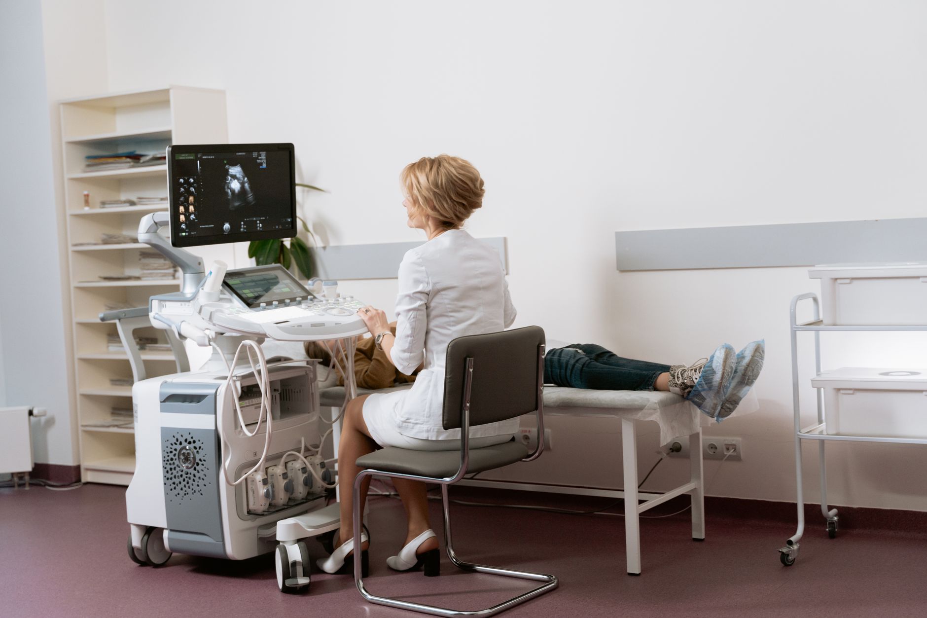
During the scan: You’ll lie on a padded table while the sonographer applies a water‑based gel to your abdomen (or gently inserts a thin probe for a transvaginal scan). The gel helps conduct sound waves. The probe moves gently across your skin, sending high‑frequency sound waves that bounce back to create the image you see on the monitor.
It’s normal to feel a slight coldness from the gel and a mild pressure from the probe. If you’re uncomfortable, let the technician know—adjustments can be made.
After the scan: You’ll hear the baby’s heartbeat and may see a tiny silhouette on the screen. The sonographer will point out key findings, such as the CRL measurement, heart rate, and NT value. If anything requires follow‑up, they’ll note it in the report and discuss next steps with you.
Some clinics provide a printed “snapshot” of the image for you to keep. While it’s tempting to hold onto every picture, remember that the clinical report—not the photo—is what matters for your medical record.
Are 12‑week pregnancy scans safe for my baby?
Yes. Ultrasound uses sound waves, not ionizing radiation, and has been used for decades without evidence of harm when performed by qualified professionals. The FDA and ACOG both state that diagnostic ultrasound is considered safe when used appropriately.
Guidelines recommend keeping the “thermal index” (a measure of energy delivered) as low as reasonably achievable. Most routine scans, including the 12‑week dating scan, fall well within these safety margins. There is no known link between diagnostic ultrasound and adverse fetal outcomes.
While the technology is safe, it’s still a medical procedure. Unnecessary “keepsake” scans are discouraged because prolonged exposure without clinical indication could theoretically increase exposure without benefit.
Professional societies such as the International Society of Ultrasound in Obstetrics and Gynecology (ISUOG) advise that each scan should have a clear clinical purpose. This principle protects both the fetus and the mother from unnecessary procedures.
What happens if the 12‑week scan reveals potential issues?
If the sonographer notes an abnormal finding—such as a significantly increased NT measurement, a missing heartbeat, or an unusual anatomy—your provider will discuss next steps. Common pathways include:
Repeat ultrasound: A follow‑up scan in a week or two can confirm whether the finding persists.
Blood tests: Non‑invasive prenatal testing (NIPT) can assess chromosomal risk using maternal blood.
Referral to a maternal‑fetal medicine specialist: For detailed counseling and possible invasive testing (e.g., chorionic villus sampling).
Most “abnormal” findings at 12 weeks turn out to be benign variants or technical artifacts. The key is early detection, which allows you and your care team to plan appropriate monitoring and support.
When a high NT measurement is detected, the combined risk algorithm (NT + serum markers) is applied. If the calculated risk exceeds a certain threshold (often 1:300), providers may recommend NIPT as a next, less invasive step before considering diagnostic testing.
Importantly, receiving an abnormal result does not mean you will have a problem; it simply means more information is needed to clarify the situation.
Additional common questions about the 12‑week scan
12 week ultrasound gender prediction accuracy
At 12 weeks, the genital tubercle is still developing, making gender identification highly unreliable. Most clinicians advise waiting until the 18‑20‑week anatomy scan, where accuracy rises to about 95 %.
12 week ultrasound heartbeat rate normal
A normal fetal heart rate at 12 weeks ranges from 120 to 180 beats per minute. Rates outside this range may warrant closer monitoring, but occasional fluctuations are common.
What to do before 12 week scan
Check with your clinic about bladder instructions, avoid heavy meals that could cause discomfort, and bring a list of any medications you’re taking. Wear comfortable clothing and consider bringing a support person if you’d like company.
12 week scan cost without insurance
In the United States, out‑of‑pocket costs vary widely, typically ranging from $150 to $300 for a standard dating scan. In the UK, NHS patients receive the scan free of charge, while private clinics may charge £100‑£200.
Difference between 12 week scan and 20 week scan
Feature
12‑week (dating) scan
20‑week (anatomy) scan
Primary purpose
Confirm viability, date pregnancy, early screening
Detailed anatomy, detect structural anomalies
Typical gestational age
11‑13 weeks
18‑22 weeks
Key measurements
Crown‑rump length, nuchal translucency
Head circumference, abdominal circumference, femur length
Gender visibility
Very limited, not reliable
Usually clear
Duration
15‑30 minutes
30‑45 minutes
Can you see baby's face at 12 weeks?
Facial features are still forming. You may see the outline of the head and sometimes the eyes as dark spots, but a recognizable face typically appears after 13‑14 weeks.
12 week ultrasound for multiples
For twins or higher‑order multiples, the scan confirms the number of embryos, assesses each heartbeat, and measures individual CRL and NT values. The procedure may take a few extra minutes, and a transvaginal approach is often used for better visualization.
What is a normal nuchal translucency measurement at 12 weeks?
Normal NT values vary by gestational age but are generally less than 2.5 mm for a 12‑week fetus. Values above 3.5 mm are considered elevated and may prompt further testing.
Transabdominal vs. transvaginal ultrasound: which is right for you?
Both approaches use the same sound‑wave technology, but they differ in how the probe reaches the uterus. A transabdominal scan is performed over a partially full bladder, which lifts the uterus for a clearer view. It’s the most common method for a first‑trimester dating scan and feels similar to a routine physical exam.
When a transabdominal view is limited—often because of a higher BMI, a retroverted uterus, or early gestational age—a transvaginal scan may be recommended. The thin probe is gently inserted into the vagina, providing closer proximity to the embryo and often sharper images. The procedure is brief, usually lasting only a few minutes, and most patients describe it as only slightly uncomfortable.
Both techniques are endorsed by ACOG and ISUOG as safe when performed by trained providers. Your clinician will choose the method that yields the best image quality while keeping your comfort in mind. If you have concerns, discuss them ahead of time; many clinics will let you choose after a brief explanation of each option.
Interpreting the fetal heartbeat rate: what’s normal and when to worry
The fetal heart rate (FHR) at 12 weeks typically falls between 120 and 180 beats per minute. A steady rate within this range is a reassuring sign of healthy cardiac development. Slight variations from beat to beat are normal and often reflect fetal movements or maternal breathing.
If the heartbeat is consistently below 110 bpm or above 190 bpm, your provider may schedule a follow‑up scan to rule out arrhythmias or other cardiac concerns. In most cases, a single out‑of‑range reading is not alarming, but persistent abnormalities warrant closer monitoring and possibly referral to a fetal cardiology specialist.
It’s also worth noting that heart rate can be temporarily affected by maternal factors such as fever, stress, or certain medications. Always share any recent illnesses or new prescriptions with your care team so they can interpret the FHR in context.
Understanding your ultrasound report: what the numbers mean
After your scan, the sonographer sends a detailed report to your obstetrician. The report typically includes the CRL measurement (in millimeters), the fetal heart rate, the NT thickness, and a qualitative note about the placenta and amniotic fluid. These numbers are compared against standardized reference ranges—often displayed as percentile charts—to assess whether the fetus is growing as expected.
If the CRL falls within the 10th‑90th percentile for the gestational age, it’s considered normal. A measurement outside this range may signal a growth restriction or a dating discrepancy, prompting the provider to reassess the estimated due date. The NT measurement is interpreted alongside maternal age and serum markers (if a combined test was performed) to generate a risk score for chromosomal abnormalities.
Most reports also include a “impression” section that succinctly states whether the findings are within normal limits or if any follow‑up is recommended. Look for phrases like “no abnormal findings” or “NT measurement elevated, recommend NIPT.” If you see medical jargon, don’t hesitate to ask your provider to clarify—understanding the report empowers you to make informed decisions.
Key measurements on the report help your provider assess growth and risk.
Special considerations: BMI, uterine fibroids, and early pregnancy challenges
Women with higher body mass index (BMI) sometimes face slightly reduced image quality because excess tissue can attenuate the ultrasound beam. In such cases, a transvaginal approach often yields clearer images, and the sonographer may need a few extra minutes to obtain the necessary measurements. The American College of Radiology acknowledges that BMI can affect visualization but emphasizes that a skilled sonographer can still acquire reliable data.
Uterine fibroids—benign growths in the uterine wall—can also cast shadows that obscure the embryo. If fibroids are present, the sonographer may adjust the probe angle or use a different scanning window to get a clear view. While most fibroids are harmless, large or rapidly growing fibroids are monitored because they can occasionally impact placental placement or cause discomfort later in pregnancy.
Other early‑pregnancy challenges, such as a tilted uterus (retroverted) or a very early gestational sac, may require the technician to use a slightly different technique. The key takeaway is that variations in anatomy rarely prevent a successful 12‑week scan; they simply mean the sonographer will adapt the exam to capture the needed images.
If you have concerns about these factors, discuss them with your provider before the appointment. Knowing ahead of time can help you plan—perhaps scheduling a transvaginal scan or allowing a little extra time for the exam.
Myth vs. fact
Myth: The 12‑week ultrasound can tell you the baby’s gender with certainty.
Fact: Gender determination is not reliable until the anatomy scan at 18‑20 weeks, when the genitalia are fully formed.
Myth: Ultrasound can harm the baby if done early.
Fact: Diagnostic ultrasound, performed by qualified professionals, has no known adverse effects on fetal development.
Myth: If the 12‑week scan looks perfect, there’s no need for any further testing.
Fact: The 12‑week scan is a screening tool; additional tests (e.g., blood work, later anatomy scan) are still recommended for comprehensive prenatal care.
Key takeaways
The 12‑week scan primarily confirms viability, refines your due date, and screens with nuchal translucency.
Typical scan time is 15‑30 minutes; preparation may involve a partially full bladder.
Heart rate of 120‑180 bpm and NT < 2.5 mm are considered normal.
Gender and detailed facial features are usually not visible until 18‑20 weeks.
Ultrasound is safe when performed by trained providers; there’s no evidence of harm.
If irregularities appear, follow‑up testing and specialist referral are standard next steps.
Understanding the written report helps you stay informed about growth and risk.
Higher BMI, fibroids, or a tilted uterus may require a transvaginal approach but rarely prevent a successful scan.
Both transabdominal and transvaginal methods are safe; your provider will choose the one that gives the best image.
Fetal heartbeat rates that stay within 120‑180 bpm are reassuring; persistent outliers warrant a repeat exam.
Frequently asked questions
What is the main purpose of the 12‑week scan?
The main purpose is to confirm that the pregnancy is viable, accurately date the pregnancy using crown‑rump length, and perform early screening such as nuchal translucency for chromosomal risk.
Can you see the baby clearly at 12 weeks?
You’ll see a tiny silhouette with a visible heartbeat, but detailed facial features are not yet clear; most parents can’t see a distinct face until around 13‑14 weeks.
What measurements are taken at the 12‑week scan?
Key measurements include crown‑rump length, heart rate, nuchal translucency thickness, and sometimes early limb or organ measurements for baseline reference.
Is a 12‑week scan mandatory?
While not legally required, most obstetric guidelines (ACOG, NICE) recommend a dating scan in the first trimester to ensure accurate dating and early assessment of pregnancy health.
How accurate is the dating scan at 12 weeks?
Dating based on crown‑rump length at 12 weeks is accurate within ±5 days for most pregnancies, making it the most reliable method for estimating the due date.
What if the baby is not visible at 12 weeks?
If the embryo isn’t visualized, the sonographer may repeat the scan in a week or two, use a transvaginal approach, or assess for possible early miscarriage. Most cases resolve with a follow‑up scan.
Can I have a 12‑week scan if I’ve had a previous miscarriage?
Yes. A prior miscarriage does not affect the safety or usefulness of the 12‑week scan. In fact, many providers use the early scan to reassure patients and establish a clear baseline after a loss.
What should I do if I hear my baby’s heartbeat before the scheduled scan?
Hearing a fetal heartbeat with a home Doppler can be reassuring, but it’s not a substitute for a professional ultrasound. Bring the information to your provider; they may note it in your chart but will still perform the standard scan to capture accurate measurements.
Can a 12‑week scan detect an ectopic pregnancy?
A 12‑week scan can sometimes identify an ectopic pregnancy if the embryo is located outside the uterus, but earlier scans (6‑10 weeks) are more reliable for this purpose. If you have pain or bleeding, your provider may order a transvaginal scan sooner.
Are there any foods or activities I should avoid before the scan?
Generally, there are no strict restrictions. However, it’s wise to avoid heavy meals right before the appointment if you’ll need a partially full bladder, and limit caffeine if you’re prone to bladder urgency. Gentle movement is fine; just follow any specific instructions your clinic gives you.
From our medical team: The 12‑week scan is a routine, low‑risk part of prenatal care. If you’re nervous, remember that the sonographer is trained to capture the images quickly and gently. Bring any questions you have, and don’t hesitate to ask for clarification on the report—your peace of mind is just as important as the data gathered.
When to call your doctor
If you notice any of the following, contact your provider promptly: no heartbeat detected, severe abdominal pain or bleeding, fever over 100.4 °F (38 °C), or if the ultrasound report mentions abnormal findings such such as a high nuchal translucency measurement.
This article provides general information and is not a substitute for personalized medical advice. Always discuss your specific situation with your obstetrician or midwife.
References
American College of Obstetricians and Gynecologists (ACOG). “Ultrasound Guidance for Pregnancy.” Practice Bulletin No. 226, 2022.
National Institute for Health and Care Excellence (NICE). “Antenatal Care for Healthy Women and Their Babies.” NG121, 2021.
U.S. Food and Drug Administration (FDA). “Diagnostic Ultrasound Safety.” Updated 2023.
World Health Organization (WHO). “Recommendations on Antenatal Care for a Positive Pregnancy Experience.” 2022.
Royal College of Obstetricians and Gynaecologists (RCOG). “Fetal Nuchal Translucency Screening.” Green‑top Guideline No. 69, 2020.
Centers for Disease Control and Prevention (CDC). “Prenatal Care and Screening.” 2023.
Mayo Clinic. “First trimester ultrasound: What to expect.” 2023.
National Health Service (NHS). “Ultrasound scans in pregnancy.” 2022.
International Society of Ultrasound in Obstetrics and Gynecology (ISUOG). “Safety and Use of Ultrasound in Pregnancy.” 2021.
BMJ. “Combined first‑trimester screening for Down syndrome: systematic review.” 2022.
American College of Radiology (ACR). “ACR–SPR Practice Parameter for Imaging in Pregnancy.” 2020.
International Society of Ultrasound in Obstetrics and Gynecology (ISUOG). “Guidelines for First‑Trimester Ultrasound.” 2021.
Editor's pick for this topic
About the Author
When Shubhra Mishra was expecting her first child in 2016, she was overwhelmed by conflicting food advice — one site said yes, another said never. By the time her second baby arrived in 2019, she realized millions of mothers face the same confusion.
That sparked a five-year journey through clinical nutrition papers, cultural diets, and expert conversations — all leading to BumpBites: a calm, compassionate space where science meets everyday motherhood.
Her long-term vision is to build a global community ensuring safe, supported, and free deliveriesfor every mother — because no woman should face pregnancy alone or uninformed. 🌿
🌍 Stand with mothers, shape safer guidance
Join a small circle of experts who review BumpBites articles so expecting parents everywhere can decide with confidence.