The FMH assessment for trauma in pregnancy uses bedside ultrasound and fetal monitoring, then follows a management protocol to protect mother and baby's safety.
By Shubhra Mishra — a mom of two who turned her own confusion during pregnancy into BumpBites, a global mission to make food choices clear, safe, and stress-free for every expecting mother. 💛
Check whether any food is safe during pregnancy with the BumpBites Food Safety Checker.
Download the Complete Pregnancy Food Guide (10,000 Foods) 📘
Instant PDF download • No spam • Trusted by thousands of moms
💡 Your email is 100% safe — no spam ever.
Quick take: When a pregnant woman experiences blunt trauma, fetal‑maternal hemorrhage (FMH) can occur silently but carries a real risk to the baby and to the mother’s Rh status. The first priority is maternal stabilization, followed by rapid FMH assessment—usually with a Kleihauer‑Betke or flow‑cytometry test—and timely Rh immunoglobulin if indicated. A step‑by‑step protocol guides monitoring, interventions, and delivery decisions, keeping both mother and fetus safe.
It’s the middle of the night, you’ve just been in a minor car accident, and the ambulance’s lights are flashing outside your bedroom window. Your heart is racing, but the paramedics reassure you that you’re stable. The next question that pops into your mind is: “Did my baby just lose blood?” You’re not alone—many expecting parents wonder about fetal‑maternal hemorrhage (FMH) after any kind of trauma.
🔢 Calculate it for your situation: Use our Kleihauer-Betke / FMH for a personalized result in seconds.
In this guide we walk you through exactly what FMH is, why it matters after a fall, a fender‑bender, or any blunt injury, and how hospitals evaluate and treat it. We’ll break down the laboratory tests, the timing of Rh immunoglobulin, and the monitoring steps that keep your baby’s heart rate under watch. By the end you’ll know the key red flags, the practical steps to expect in the emergency department, and the follow‑up care you’ll need after you leave the hospital.
We’ll also provide a quick reference table, a handy calculator link for estimating blood loss, and a myth‑busting section so you can separate fact from fiction. Everything is rooted in guidance from ACOG, RCOG, the CDC, NHS, and other leading bodies, and we’ve included a doctor’s note for that extra reassurance.
What is fetal‑maternal hemorrhage (FMH) and why trauma matters?
Fetal‑maternal hemorrhage describes the passage of fetal red blood cells (RBCs) into the maternal circulation. In a healthy pregnancy a tiny amount—often less than 0.5 mL—crosses the placenta daily without consequence. After blunt trauma, especially in the second or third trimester, the placenta can be disrupted, leading to a sudden surge of fetal blood into the mother’s bloodstream.
Why does this matter? First, a large FMH can cause fetal anemia, which may progress to fetal distress, growth restriction, or even stillbirth if not recognized. Second, if the mother is Rh‑negative and the fetus is Rh‑positive, the fetal cells can trigger an immune response that leads to alloimmunization. This sensitization dangerously endangers any future pregnancies, because the mother’s antibodies can cross the placenta and attack subsequent Rh‑positive fetuses.
Overall, the risk of clinically significant FMH after trauma is low—studies from the American College of Surgeons Trauma Quality Improvement Program report rates of 1–2 % in major blunt injuries—but the consequences are serious enough that protocols exist to catch it early. The NHS also notes that while most minor injuries do not cause FMH, clinicians remain vigilant because the placenta’s vascular network is highly sensitive to sudden force. The mechanism often involves shear forces, where the sudden deceleration or impact causes the placenta to detach slightly from the uterine wall, tearing delicate fetal vessels.
Initial assessment and stabilization of a pregnant trauma patient
The f
irst minutes follow the same algorithm as any trauma patient: ABCs—airway, breathing, circulation. The presence of a pregnancy does not change the need to secure the airway or control bleeding, but it does add layers of consideration.
Airway and breathing: Position the patient in a left‑lateral tilt (or use a wedge) to relieve aortocaval compression, especially after the 20‑week mark. Supplemental oxygen should be given if saturation falls below 94 %.
Circulation: Assess pulse, blood pressure, and capillary refill. Intravenous access (two large‑bore lines) is obtained immediately; fluids are given judiciously to avoid uterine hypervolemia that could worsen placental separation.
Disability and exposure: A rapid neurologic check (Glasgow Coma Scale) is performed, and the abdomen is examined for tenderness, bruising, or signs of uterine rupture.
Obstetric assessment: Once the mother is stable, a bedside obstetric ultrasound is ordered to confirm fetal viability, gestational age, and placental location.
Maternal stabilization is the gateway to FMH evaluation—if the mother is hypotensive or bleeding heavily, addressing those needs takes precedence. However, the trauma team should simultaneously flag the case for obstetric consultation because fetal monitoring may need to start within minutes. ACOG’s “Trauma in Pregnancy” bulletin stresses that early obstetric involvement reduces the chance of missed FMH and improves neonatal outcomes. It’s also important to remember that physiological changes in pregnancy, such as increased blood volume and heart rate, can mask signs of shock, making vigilance even more critical.
Diagnosing FMH: Kleihauer‑Betke, flow cytometry, and other tools
After the initial stabilization, the next step is to quantify any fetal blood that has entered the maternal circulation. The two most common laboratory methods are the Kleihauer‑Betke (KB) test and flow cytometry.
The Kleihauer‑Betke test is a microscopy‑based assay that exploits the fact that fetal hemoglobin (HbF) is resistant to an acid‑elution process that washes out adult hemoglobin (HbA). After staining, fetal cells appear bright pink against a pale background of maternal cells. The lab counts the number of stained cells per 5,000–10,000 maternal cells, allowing calculation of the volume of FMH. The test is inexpensive, widely available, and can be performed on a standard blood sample, but it is operator‑dependent and may underestimate FMH when the fetal cells are fragmented.
In contrast, flow cytometry uses fluorescently labeled antibodies that bind specifically to HbF or fetal‑specific antigens (e.g., CD45). The machine counts millions of cells, providing a highly sensitive and precise estimate of FMH, often down to 0.01 % of total maternal blood. The downside is that it requires specialized equipment and may not be accessible in all emergency departments.
Both tests have a turnaround time of 4–6 hours in most hospitals, which is fast enough for the acute management window. When the initial KB result is borderline, many centers reflexively send a sample for flow cytometry to confirm the volume. The CDC’s guidance on Rh immunoglobulin administration recommends that labs prioritize STAT processing for pregnant trauma patients. Proper sample collection and handling are crucial for accurate results, and any delays can impact timely intervention. While awaiting definitive FMH results, indirect signs like abnormal fetal Doppler studies might raise initial concern for fetal anemia.
For clinicians who need to calculate the exact amount of fetal blood to determine Rh immunoglobulin dosing, the Kleihauer‑Betke / FMH calculator can be a handy bedside tool.
Feature
Kleihauer‑Betke
Flow Cytometry
Principle
Acid elution & staining of HbF
Fluorescent antibodies targeting HbF
Sensitivity
Detects ≥0.5 % FMH
Detects ≥0.01 % FMH
Turn‑around time
4–6 hours (standard lab)
6–8 hours (requires specialized lab)
Availability
Most hospitals
Limited to tertiary centers
Cost
Low
Higher
Operator dependence
High
Low
Technicians use the Kleihauer‑Betke test to spot fetal cells that have entered the mother’s bloodstream.
Rh immunoglobulin: when, how much, and dosing criteria
Rh immunoglobulin (RhIg) is a pooled IgG preparation that masks fetal Rh‑positive red cells, preventing the mother’s immune system from recognizing them. The CDC and ACOG recommend RhIg whenever a pregnant Rh‑negative woman is at risk of fetal‑blood exposure, which includes any trauma that could cause FMH.
Guidelines for dosing are straightforward: the standard dose (300 µg, often labeled as “250 µg anti‑D”) covers up to 30 mL of fetal whole blood. If the calculated FMH exceeds 30 mL, additional doses are given in proportion to the volume. For example, a KB result indicating 60 mL of FMH would require two standard doses. The NHS also notes that each 300 µg dose is equivalent to 0.5 mL of Rh‑positive red cells, providing a clear conversion for clinicians.
Timing matters. RhIg should be administered within 72 hours of the injury to be most effective. If the exact FMH volume is not yet known, many providers give a prophylactic dose immediately and adjust later if the lab confirms a larger volume. This “early‑and‑often” approach aligns with ACOG’s 2022 practice bulletin, which stresses that delayed administration reduces efficacy and increases the chance of sensitization. Rest assured, RhIg is a highly purified blood product with a long track record of safety, and any mild side effects like injection site soreness are far outweighed by its critical role in preventing Rh sensitization.
Special circumstances—such as multiple injuries, ongoing bleeding, or a need for emergency cesarean—may prompt repeat dosing. ACOG also advises a follow‑up antibody screen at 28 weeks and again at delivery to ensure no sensitization has occurred.
Understanding the Rh Factor: A Quick Primer for Expecting Parents
You may have heard your doctor mention your "Rh factor" during your first prenatal visit. This refers to a specific protein found on the surface of red blood cells. If you have the protein, you're Rh-positive; if you don't, you're Rh-negative. Most people are Rh-positive, but about 15% of the population is Rh-negative.
For most of life, your Rh status doesn't matter. But in pregnancy, if an Rh-negative mother carries an Rh-positive baby, her immune system can recognize the baby's Rh-positive red blood cells as "foreign." This can happen if even a small amount of fetal blood enters her circulation, as in a trauma. Her body then creates antibodies against these cells. While this usually doesn't affect the current pregnancy, these antibodies can attack the red blood cells of a future Rh-positive baby, leading to serious complications like severe anemia, jaundice, and even fetal hydrops, a life-threatening condition. This is why Rh immunoglobulin (RhIg) is so crucial: it "hides" any fetal Rh-positive cells from the mother's immune system, preventing her from developing those antibodies. (ACOG, CDC guidance).
Step‑by‑step management protocol after suspected FMH
Maternal stabilization: Follow ABCs, left‑lateral tilt, IV access, and pain control. Document vital signs every 15 minutes.
Obstetric consultation: Notify the obstetric team as soon as the patient is stable enough for a brief exam. They will order bedside ultrasound to confirm fetal heart rate (FHR) and viability.
Fetal monitoring: Initiate continuous electronic fetal monitoring (EFM) if the gestation is ≥24 weeks. Look for baseline rate 110–160 bpm, variability, and decelerations. Any non‑reassuring pattern warrants immediate evaluation.
Blood sampling for FMH: Draw maternal peripheral blood for KB and, if available, flow cytometry. Send the sample with a “STAT” tag.
RhIg administration: If the mother is Rh‑negative, give a standard 300 µg dose within 72 hours, even before lab results if the injury was significant (e.g., abdominal impact, >5 cm bruising over uterus).
Quantify FMH: Once the KB result is back, calculate the volume using the formula:
FMH (mL) = [(Number of fetal cells / Number of maternal cells) × Maternal blood volume] ÷ 100.
Maternal blood volume is estimated at 5,000 mL for a 70‑kg adult; adjust for body weight if known.
Additional RhIg dosing: If FMH > 30 mL, give an extra 300 µg dose for each additional 30 mL (or proportionally less if fractional).
Decide on delivery: If the fetus shows persistent distress, severe anemia (evidenced by Doppler studies or loss of variability), or placental abruption, delivery may be indicated. The mode (vaginal vs. cesarean) follows standard obstetric criteria and gestational age.
Post‑delivery care: Test the neonate’s blood type and direct Coombs test. If the baby is Rh‑positive, give prophylactic RhIg to the newborn as per neonatal guidelines.
Follow‑up: Schedule a maternal antibody screen at 28 weeks and a repeat test 4 weeks after any subsequent trauma. Provide counseling on signs of anemia and encourage regular prenatal visits.
Throughout the protocol, communication between the trauma team, obstetrics, and the laboratory is critical. A clear, written checklist (often kept in the ED’s “Pregnancy Trauma” binder) helps ensure no step is missed. ACOG’s 2022 bulletin even provides a printable flowchart that many hospitals have adopted. This protocol is a dynamic framework, requiring continuous reassessment of both maternal and fetal status. The team must be prepared to escalate care quickly if conditions change, always prioritizing the well-being of both mother and baby.
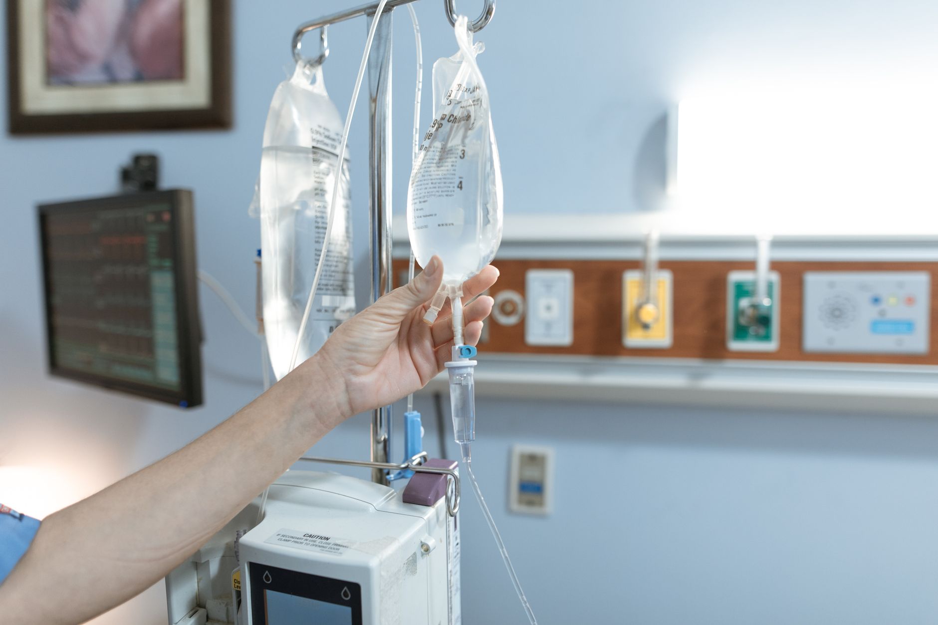
Continuous fetal monitoring is started as soon as the mother’s condition allows.
The Role of Ultrasound in Trauma and FMH Evaluation
Beyond simply confirming fetal viability, ultrasound plays a crucial role in the comprehensive evaluation of a pregnant trauma patient. A bedside ultrasound, often performed by the obstetric team, can rapidly assess several key factors. It can determine gestational age, confirm fetal heart activity, and crucially, evaluate the placenta for signs of abruption, such as a retroplacental clot or changes in uterine wall integrity. (SMFM Clinical Guidance, 2023).
Ultrasound also allows for the assessment of amniotic fluid volume and can identify any free fluid in the maternal abdomen, which could indicate internal bleeding. In cases of suspected FMH, serial ultrasounds may be used to monitor fetal growth and look for signs of fetal anemia, such as increased velocity in the fetal middle cerebral artery. This comprehensive imaging helps guide management decisions, from whether to continue monitoring to the timing and mode of delivery.
Guidelines for fetal monitoring, delivery decisions, and obstetric consultation after trauma
Fetal monitoring after trauma follows the same thresholds as any other high‑risk pregnancy. The American College of Obstetricians and Gynecologists (ACOG) advises:
Continuous EFM for at least 4 hours after a significant blunt injury if the gestation is ≥24 weeks.
If the FHR pattern shows late decelerations, absent variability, or bradycardia (<110 bpm), immediate obstetric evaluation is required.
Placental location and integrity should be assessed via transabdominal ultrasound; signs of abruption (retroplacental clot, increased uterine tone) may prompt early delivery.
When delivery is indicated before 34 weeks, corticosteroids (betamethasone 12 mg IM × 2) are administered to mature fetal lungs, unless contraindicated by maternal instability.
Delivery mode is individualized. A stable mother with a cephalic‑presenting fetus and no obstetric contraindications may have a vaginal delivery, even after trauma. However, if there is significant uterine rupture, fetal distress, or maternal hemodynamic compromise, a cesarean section is often the safest route. The NHS notes that rapid decision‑making in these scenarios improves neonatal Apgar scores and reduces NICU admissions.
Obstetric consultation should be documented in the chart with a clear plan: monitoring schedule, thresholds for escalation, and a timeline for repeat imaging. Many hospitals have a “Pregnancy Trauma” protocol that includes a pre‑printed order set to streamline this process. Reassuring fetal heart rate patterns typically show a baseline between 110-160 bpm with good variability (fluctuations of 6-25 bpm) and accelerations. Non-reassuring patterns like late decelerations (dips in heart rate after the peak of a contraction) can signal fetal compromise and require urgent attention. (ACOG Practice Bulletin No. 232).
Risk factors, preventive measures, and postpartum follow‑up care
Not all trauma leads to FMH, but certain factors increase the likelihood:
Gestational age ≥ 20 weeks (the placenta is larger and more vascular).
Direct abdominal impact, especially from a motor vehicle accident (seatbelt injuries) or falls from a height.
Placental abnormalities (previa, low‑lying placenta) that make the uterine wall more vulnerable.
Maternal hypertension or pre‑eclampsia, which can predispose to placental separation.
Preventive strategies focus on safety: using seatbelts correctly (lap belt low on the hips, shoulder belt across the chest), avoiding high‑risk activities, and ensuring a safe home environment (non‑slippery floors, handrails on stairs). Prenatal visits should include counseling on these topics, especially in the second trimester when mobility increases. Moreover, healthcare providers are increasingly vigilant in screening for domestic violence, which is a significant, often hidden, cause of trauma in pregnancy. Offering resources and support in a trauma-informed way is a crucial part of comprehensive prenatal care. (NICE Clinical Knowledge Summary, 2022).
After the acute episode, postpartum care includes:
Repeat maternal blood type and antibody screen at 6 weeks postpartum.
Neonatal testing for hemolysis if FMH was significant, including a bilirubin level and direct Coombs test.
Psychological support if the trauma was severe; many parents experience anxiety or post‑traumatic stress, and referral to counseling can be beneficial.
Documentation of the incident in the obstetric record so future providers are aware of the Rh immunoglobulin history.
Placental abruption and FMH: a closer look
Placental abruption—premature separation of the placenta from the uterine wall—is one of the most serious complications that can accompany FMH after trauma. When the placenta detaches, fetal blood vessels can rupture, allowing a larger volume of fetal blood to spill into the maternal circulation. ACOG’s 2021 guideline notes that abruptio placentae after blunt trauma occurs in roughly 5 % of severe cases, but the risk rises dramatically with high‑velocity impacts.
Clinically, abruption may present with abdominal pain, uterine tenderness, and vaginal bleeding, though some women experience only subtle signs. Ultrasound can sometimes visualize a retroplacental clot, but the definitive diagnosis often rests on clinical suspicion and the presence of fetal distress. When abruption is confirmed, the management algorithm accelerates: immediate delivery is recommended if the fetus is viable and the mother’s condition allows, and RhIg dosing follows the same principles as for isolated FMH. The severity of abruption can vary from partial to complete separation, directly impacting the volume of FMH and the urgency of intervention. Concealed abruption, where bleeding is trapped internally, can be particularly challenging to diagnose, relying heavily on clinical signs like persistent abdominal pain, uterine rigidity, and non-reassuring fetal heart patterns. (NHS Clinical Knowledge, 2023).
Psychological support and coping after pregnancy trauma
Even when the physical injuries are minor, the emotional impact of a trauma during pregnancy can be profound. Anxiety about the baby’s health, intrusive thoughts about the accident, and fear of future injuries are common. The NHS’s perinatal mental health guidelines advise that providers screen for post‑traumatic stress disorder (PTSD) at the postpartum visit, especially after a significant incident.
Practical coping strategies include: keeping a symptom diary, leaning on a trusted support person, and practicing gentle relaxation techniques such as guided breathing or prenatal yoga (as long as it’s approved by the provider). If anxiety interferes with daily life, a referral to a perinatal therapist or a community mental‑health service is appropriate. Early intervention can prevent chronic mood disorders and improve bonding with the newborn. Therapeutic approaches like Eye Movement Desensitization and Reprocessing (EMDR) or Cognitive Behavioral Therapy (CBT) can be particularly effective for processing trauma. Remember, seeking help is a sign of strength and can significantly improve your overall well-being and ability to enjoy your pregnancy and new parenthood.
What to Expect During Your Hospital Stay and Discharge Planning
After a trauma, your hospital stay will vary depending on the severity of the injury and the findings from your assessments. For most minor traumas, continuous fetal monitoring will typically last at least 4 hours, sometimes up to 24 hours, to ensure there are no delayed signs of distress or placental issues. During this time, nurses and doctors will frequently check your vital signs, pain levels, and the baby's heart rate. You'll likely undergo the FMH blood tests, and if you're Rh-negative, you'll receive your RhIg injection.
Before discharge, your medical team will confirm that both you and your baby are stable, with no signs of ongoing bleeding, distress, or complications. You'll receive clear instructions on what signs to watch for at home, such as new abdominal pain, vaginal bleeding, fluid leakage, or changes in fetal movement. It’s crucial to attend all follow-up appointments, including repeat antibody screens if you're Rh-negative, and to communicate any lingering concerns with your obstetric provider. Don't hesitate to ask questions about activity restrictions, medication use, or any emotional support you might need.
From our medical team: While trauma during pregnancy can be incredibly frightening, modern medical protocols are designed to protect both mother and baby. Our primary goal is always maternal stabilization, followed by meticulous fetal assessment and timely interventions like Rh immunoglobulin. Remember that many women experience trauma during pregnancy and go on to have healthy babies, thanks to these comprehensive care pathways. Trust your healthcare team and don't hesitate to voice any concerns.
🔢 Ready to crunch your numbers? Use our Kleihauer-Betke / FMH for a personalized result in seconds.
Myth vs. fact
Myth: If I feel fine after a car crash, my baby is definitely safe.
Fact: The mother can be asymptomatic while the fetus loses blood. A brief evaluation, including fetal monitoring and possibly a Kleihauer‑Betke test, is recommended for any blunt trauma after the first trimester.
Myth: Only severe injuries cause FMH.
Fact: Even low‑speed collisions can disrupt placental vessels. Guidelines advise assessment for any trauma that involves abdominal impact, regardless of severity.
Myth: Rh immunoglobulin is only needed if the baby is known to be Rh‑positive.
Fact: Because fetal blood may not be known at the time of injury, Rh‑negative mothers receive prophylactic RhIg when there is any risk of fetal‑blood exposure, including trauma.
Key takeaways
Maternal stabilization always comes first; obstetric concerns are addressed in parallel.
FMH can be quantified quickly with a Kleihauer‑Betke test; flow cytometry offers higher sensitivity when available.
Rh‑negative mothers should receive a standard 300 µg RhIg dose within 72 hours of any trauma that could expose fetal blood.
Continuous fetal monitoring for 4 hours after significant blunt injury helps catch early signs of distress.
Delivery decisions are based on fetal status, gestational age, and maternal stability—not solely on the presence of FMH.
Follow‑up antibody screens and neonatal testing are essential to ensure no sensitization or anemia has occurred.
Consider placental abruption as a possible companion to FMH, and be aware of the emotional toll that trauma can take.
Ultrasound is a crucial tool for assessing placental integrity, fetal well-being, and internal injuries after trauma.
Frequently asked questions
What is fetal‑maternal hemorrhage?
Fetal‑maternal hemorrhage (FMH) is the transfer of fetal red blood cells into the mother’s bloodstream, which can happen naturally in small amounts but may increase dramatically after trauma.
How is FMH diagnosed after trauma?
Doctors usually order a Kleihauer‑Betke test, which stains fetal cells pink, or flow cytometry, which counts cells with fluorescent antibodies; both quantify the volume of fetal blood in the maternal circulation.
When should Rh immunoglobulin be given after a car accident in pregnancy?
If the mother is Rh‑negative, a standard 300 µg dose of RhIg should be administered within 72 hours of the injury, even if the exact FMH volume is not yet known.
What are the signs of fetal distress after maternal trauma?
Non‑reassuring fetal heart rate patterns—late decelerations, loss of variability, bradycardia—or reduced fetal movement reported by the mother signal possible distress and need immediate obstetric evaluation.
Which tests are used to quantify FMH?
The Kleihauer‑Betke test and flow cytometry are the two primary methods; the former is widely available, while the latter provides greater sensitivity and precision.
What is the emergency management protocol for trauma in pregnancy?
First, stabilize the mother (ABCs, left‑lateral tilt). Then, involve obstetrics, start continuous fetal monitoring, draw blood for FMH testing, give RhIg if indicated, and decide on delivery based on fetal status and gestational age.
How long will I be monitored in the hospital after trauma?
For most minor blunt traumas, continuous fetal monitoring is recommended for at least 4 hours, and often up to 24 hours, to ensure no delayed signs of fetal distress or placental complications arise before discharge. Your medical team will determine the precise duration based on your specific situation.
What if I'm Rh-negative but my baby's father is also Rh-negative?
If both you and the baby's biological father are Rh-negative, then your baby will also be Rh-negative, and you will not need Rh immunoglobulin (RhIg) after trauma or at any other time during pregnancy, as there is no risk of Rh sensitization. However, your provider will still confirm both parents' Rh status or assume Rh-positive fetal blood if paternal status is unknown.
Can I use over‑the‑counter pain relievers after a trauma?
Acetaminophen is generally considered safe in pregnancy and can be used for mild pain, but NSAIDs such as ibuprofen are avoided after 20 weeks because they may affect fetal kidney function. Always check with your provider before taking any medication.
Is it safe to travel by car after a minor accident?
Most women can resume driving once they feel comfortable and any pain is controlled. However, if you experience lingering abdominal tenderness, vaginal bleeding, or changes in fetal movement, you should seek medical evaluation before traveling long distances.
When to call your doctor
If you experience any of the following after a trauma, contact your obstetric provider or go to the nearest emergency department immediately: severe abdominal pain, vaginal bleeding, loss of fetal movement, persistent uterine cramps, signs of shock (fast heartbeat, dizziness), or any abnormal fetal heart rate reported by monitoring equipment.
This article is for informational purposes only and does not replace personalized medical advice. Always discuss your specific situation with your healthcare provider.
References
American College of Obstetricians and Gynecologists. “Management of Trauma in Pregnancy.” ACOG Practice Bulletin No. 232, 2022.
Royal College of Obstetricians and Gynaecologists. “The Management of Fetal‑Maternal Hemorrhage.” RCOG Green‑top Guideline, 2021.
Centers for Disease Control and Prevention. “Rh Immunoglobulin (RhIg) Recommendations for Pregnant Women.” CDC Guidelines, 2023.
National Institute for Health and Care Excellence. “Pregnancy and Trauma.” NICE Clinical Knowledge Summary, 2022.
World Health Organization. “Maternal and Perinatal Health: Guidelines for Managing Trauma in Pregnancy.” WHO Publication, 2021.
Mayo Clinic. “Kleihauer‑Betke Test.” Mayo Clinic Proceedings, 2020.
Society for Maternal‑Fetal Medicine. “Fetal Monitoring after Maternal Trauma.” SMFM Clinical Guidance, 2023.
American Red Cross. “Emergency Management of Pregnant Trauma Patients.” Red Cross Protocol, 2022.
British Columbia Ministry of Health. “Rh Immunoglobulin Administration after Trauma.” Provincial Health Guidelines, 2022.
National Perinatal Information Center. “Post‑Trauma Follow‑Up for Pregnant Women.” NPIC Report, 2021.
National Health Service (NHS). “Placental Abruption.” NHS Clinical Knowledge, 2023.
American College of Surgeons. “Trauma Quality Improvement Program: Pregnancy Sub‑Study.” ACS Data, 2020.
Editor's pick for this topic
About the Author
When Shubhra Mishra was expecting her first child in 2016, she was overwhelmed by conflicting food advice — one site said yes, another said never. By the time her second baby arrived in 2019, she realized millions of mothers face the same confusion.
That sparked a five-year journey through clinical nutrition papers, cultural diets, and expert conversations — all leading to BumpBites: a calm, compassionate space where science meets everyday motherhood.
Her long-term vision is to build a global community ensuring safe, supported, and free deliveriesfor every mother — because no woman should face pregnancy alone or uninformed. 🌿
🌍 Stand with mothers, shape safer guidance
Join a small circle of experts who review BumpBites articles so expecting parents everywhere can decide with confidence.