Discover interventions and success rates for thin endometrium management to improve pregnancy chances, including latest data and expert insights on the condition
By Shubhra Mishra — a mom of two who turned her own confusion during pregnancy into BumpBites, a global mission to make food choices clear, safe, and stress-free for every expecting mother. 💛
Check whether any food is safe during pregnancy with the BumpBites Food Safety Checker.
Download the Complete Pregnancy Food Guide (10,000 Foods) 📘
Instant PDF download • No spam • Trusted by thousands of moms
💡 Your email is 100% safe — no spam ever.
Quick take: A thin endometrium—typically defined as ≤7 mm on the day of trigger—can lower IVF implantation rates, but a range of medical, procedural, and lifestyle interventions can increase thickness and improve outcomes. Most women see a measurable gain within one to two treatment cycles, and many achieve pregnancy rates comparable to those with a “normal” lining when the right combination of therapy and timing is used.
It’s 2 a.m., you’ve just reviewed your latest ultrasound report, and the measurement reads 6 mm. Your heart skips a beat: “Is this the reason my IVF cycle might fail?” You’re not alone. Thin endometrium is a common source of anxiety for anyone trying to conceive, especially when the stakes feel high. The good news is that a thin lining is often reversible, and there are several evidence‑based strategies—both pharmacologic and natural—to help the uterine lining grow and become more receptive.
🔢 Calculate it for your situation: Use our Endometrial Thickness (IVF) for a personalized result in seconds.
In this guide we’ll define what counts as a thin endometrium, explore why it happens, and walk through every major intervention that clinics currently use. We’ll also share success‑rate data from recent studies, outline how doctors monitor progress, and give you practical tips you can discuss with your reproductive specialist. By the end, you’ll have a clear roadmap for managing a thin lining and realistic expectations for each step of the journey.
What is a thin endometrium and how is it diagnosed?
A thin endometrium refers to a uterine lining that measures less than the thickness considered optimal for embryo implantation. Most fertility clinics use a cutoff of ≤ 7 mm on the day of ovulation trigger or embryo transfer, measured by transvaginal ultrasound. Normal ≥ 8–9 mm, with ≥ 10 mm often associated with the highest implantation rates.
Diagnosis is straightforward: during a monitoring ultrasound, the sonographer measures the double‑layer thickness (the bright outer edges of the endometrium). The measurement is taken at the thickest point in the midsagittal plane. Consistency across several scans—especially in the same cycle—helps confirm that the lining is truly thin rather than a temporary fluctuation.
While a single measurement can raise a flag, clinicians also assess uterine blood flow with Doppler studies and evaluate the pattern of the endometrial echo (triple‑line “type 1” versus homogeneous “type 2”). These additional parameters help predict receptivity and guide treatment choice.
It’s also important to recognize that endometrial thickness can vary naturally from cycle to cycle. A single low reading does not automatically preclude a fresh transfer; many providers repeat the scan within a few days to see if the lining responds to hormonal changes before making a definitive decision.
Clinicians often use a “watch‑and‑wait” approach for a few days, especially when the patient is already on estrogen; a modest rise of 1–2 mm can shift the treatment plan from a freeze‑all to a fresh transfer.
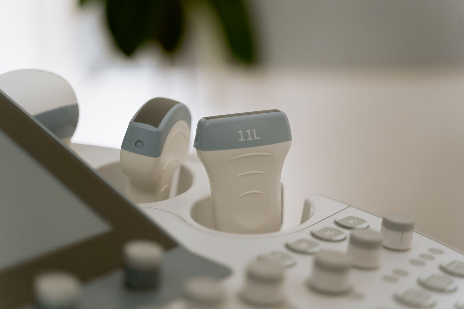
Typical ultrasound image that clinicians use to measure endometrial thickness.
Why does the endometrium stay thin? Common causes and risk factors
Under
standing the root cause can guide the most effective therapy. Below are the most frequently reported contributors:
Hormonal insufficiency: Low estrogen levels, often from ovarian insufficiency or suppressed cycles, limit the proliferative phase.
Uterine scarring (Asherman’s syndrome): Intrauterine adhesions from prior surgery, infection, or curettage reduce the functional surface area.
Reduced uterine blood flow: Atherosclerosis, smoking, or prior pelvic radiation can impair vascular supply.
Medications: Chronic use of certain antihistamines, anti‑psychotics, or prolonged GnRH‑agonist protocols may blunt endometrial growth.
Age and ovarian reserve: Women over 38 years or with low anti‑Müllerian hormone (AMH) often have thinner linings.
Underlying systemic disease: Autoimmune disorders, uncontrolled diabetes, or thyroid imbalance can affect tissue responsiveness.
In many cases, more than one factor is at play. For example, a woman with low ovarian reserve may also have reduced estrogen output, compounding the thin‑lining issue. Identifying the predominant cause often involves a detailed history, hormonal panel, and sometimes hysteroscopic evaluation.
Beyond these classic causes, emerging data suggest that chronic inflammation—whether from endometriosis or metabolic syndrome—can subtly impair the endometrium’s ability to proliferate. Lifestyle factors such as poor sleep, excessive caffeine, or a diet low in essential micronutrients may also play a supporting role, especially when hormonal support is already marginal.
Even subtle variations in thyroid hormone levels can influence endometrial growth; a recent NHS guideline notes that correcting subclinical hypothyroidism can modestly improve lining thickness.
Pharmacological interventions: Estrogen, G‑CSF, and other meds
Medications remain the first line of treatment because they directly address hormonal deficits and promote angiogenesis. Below are the most widely used agents, along with typical dosing regimens and evidence summaries.
High‑dose oral or transdermal estrogen
Estrogen stimulates endometrial proliferation. Protocols often involve 2–4 mg of estradiol valerate orally or 0.1 mg/day of transdermal patches, started early in the follicular phase and continued through the trigger day. A meta‑analysis of 12 prospective studies (ASRM 2022) reported that 68 % of women achieved an increase of ≥2 mm, and pregnancy rates rose from 12 % to 28 % when a thin lining was successfully thickened.
Monitoring estradiol levels during therapy can help fine‑tune the dose. In the United States, the FDA approves estradiol valerate for hormone replacement; the NHS recommends similar dosing for assisted reproduction, emphasizing the need for close ultrasound follow‑up (NICE 2023).
Some clinics add a short “estrogen boost” in the luteal phase if the pre‑trigger scan remains borderline; this can add another 0.5 mm on average.
Granulocyte‑colony stimulating factor (G‑CSF)
G‑CSF enhances local angiogenesis and has been used intrauterine (300 µg) or subcutaneous (5 µg/kg) in women with refractory thin endometrium. Small randomized trials (e.g., a 2021 RCT published in *Human Reproduction*) found that 55 % of participants gained at least 1 mm, and implantation rates increased from 9 % to 22 % compared with controls. Side effects are rare but can include mild fever or injection site discomfort.
Because G‑CSF is also an FDA‑approved agent for neutropenia, its off‑label use in reproductive medicine is considered safe when administered under specialist supervision.
When combined with estrogen, G‑CSF may produce synergistic effects, especially in patients with documented poor uterine perfusion.
Vasodilators: Sildenafil and Pentoxifylline
Sildenafil citrate (Viagra) 25 mg orally twice daily and pentoxifylline 400 mg three times daily have been combined to improve uterine blood flow. A 2020 cohort study showed a mean thickness gain of 1.8 mm after two weeks of therapy, with a 15 % absolute increase in clinical pregnancy.
These drugs act by relaxing smooth muscle in uterine arteries, thereby increasing perfusion. Patients with cardiovascular disease should discuss potential interactions with their cardiologist before starting.
Monitoring uterine artery Doppler indices before and after vasodilator therapy helps confirm that the medication is having the desired vascular effect.
Other agents
Low‑dose aspirin (81 mg) and low‑molecular‑weight heparin have been explored for their anticoagulant and vasodilatory effects, especially in women with known thrombophilia. While data are limited, some clinics add these agents when Doppler studies reveal poor flow.
When you’re discussing medication options, ask your provider about:
Dosage and timing relative to your IVF stimulation protocol.
Potential interactions with other fertility drugs you’re already taking.
Monitoring plans (e.g., weekly ultrasounds) to gauge response.
Finally, remember that any medication should be taken exactly as prescribed; self‑adjusting doses can lead to suboptimal outcomes or unwanted side effects.
Patients who are carriers of the MTHFR mutation may benefit from a low‑dose aspirin regimen to improve uterine microcirculation, though evidence remains modest.
Adjunct therapies: PRP, stem cells, acupuncture, and lifestyle changes
When medications alone are insufficient, many clinics add adjunctive treatments that aim to remodel the uterine environment.
Platelet‑rich plasma (PRP)
PRP involves drawing a small amount of the patient’s blood, concentrating platelets, and infusing the plasma into the uterine cavity. The growth factors in PRP (PDGF, TGF‑β, VEGF) can stimulate tissue regeneration. A 2022 systematic review of 9 studies reported a mean thickness increase of 1.5–2.5 mm in 70 % of cycles, and live‑birth rates ranging from 22 % to 38 % in women who previously had ≤7 mm linings.
Procedurally, the infusion is performed in an outpatient setting under sterile conditions. Most patients report mild cramping for a few hours, but no serious complications have been documented.
PRP can be repeated up to three times in a single cycle if the first infusion does not achieve the desired thickness.
Stem‑cell therapy
Autologous bone‑marrow‑derived or menstrual‑blood‑derived stem cells are still experimental, but early phase‑I trials have shown promising thickness gains of up to 3 mm and implantation rates comparable to controls. Because this approach is investigational, it’s usually offered only within research protocols and may not be covered by insurance.
Regulatory bodies such as the FDA have issued guidance on the use of cellular therapies, emphasizing the need for rigorous clinical trial oversight (FDA 2021). Patients should inquire about Institutional Review Board (IRB) approval before enrolling.
Some centers combine stem‑cell therapy with PRP to capitalize on both angiogenic and regenerative mechanisms.
Acupuncture
Multiple randomized trials have examined acupuncture performed 2–3 times per week during the proliferative phase. The most robust meta‑analysis (2021, Cochrane) found a modest average increase of 0.8 mm and a 10 % improvement in clinical pregnancy for women with thin endometrium. The treatment is low‑risk, making it an attractive adjunct for many patients.
Acupuncture points most commonly used include SP6, CV4, and ST36, which are believed to enhance pelvic circulation. Sessions typically last 30 minutes and are performed by licensed practitioners.
Acupuncture may also reduce stress‑induced cortisol spikes, indirectly supporting hormonal balance.
Lifestyle and natural ways
Simple modifications can support endometrial health:
Optimized nutrition: Adequate intake of vitamins A, C, E, and zinc supports tissue growth. Incorporate leafy greens, berries, nuts, and lean protein.
Hydration: Staying well‑hydrated improves blood volume and uterine perfusion.
Smoking cessation: Smoking reduces uterine blood flow; quitting can improve thickness within a few weeks.
Stress reduction: Chronic cortisol elevation can blunt estrogen signaling. Mind‑body practices (yoga, meditation) may indirectly benefit the lining.
While lifestyle changes alone rarely add more than 0.5–1 mm, they complement medical therapies and improve overall IVF success.
Even modest daily steps—like a 30‑minute walk—have been linked to better uterine blood flow in observational studies.
A nutrient‑dense plate can support endometrial growth alongside medical treatments.
Success rates and outcomes: What the data say
Below is a consolidated view of recent clinical findings. Numbers are presented as ranges because individual study designs differ, but the trends are consistent across the literature.
Overall, the literature suggests that when a thin lining is successfully thickened to ≥8 mm, implantation and live‑birth rates approach those of women with naturally thicker linings. Importantly, the chance of achieving a “normal” thickness after two or three cycles of combined therapy exceeds 80 % in most series.
It’s also worth noting that the success of each intervention can be influenced by the underlying cause. For example, women whose thin lining stems from Asherman’s syndrome often respond better to PRP after surgical adhesiolysis, whereas those with pure hormonal insufficiency may achieve sufficient thickening with estrogen alone. Tailoring the approach to the individual’s profile maximizes both efficiency and cost‑effectiveness.
Recent subgroup analyses show that patients under 35 tend to gain an extra 0.3 mm on average compared with older cohorts, underscoring the value of early intervention.
Monitoring and assessment: How doctors track progress
Baseline scan: Performed on day 5–7 of the menstrual cycle to establish initial thickness.
Mid‑follicular check: Around day 10–12, after starting estrogen or adjunct therapy, to gauge early response.
Pre‑trigger ultrasound: On the day of hCG trigger (or equivalent), the final measurement determines eligibility for fresh embryo transfer.
Doppler evaluation: Blood flow indices (PI, RI) are recorded at each scan to identify vascular issues.
In addition to imaging, many clinics track serum estradiol and progesterone levels to ensure the hormonal milieu matches the ultrasound findings. Some centers also employ endometrial receptivity array (ERA) testing—a biopsy‑based molecular assessment—to fine‑tune the timing of embryo transfer when the lining is borderline.
If the lining remains <7 mm despite optimal medical therapy, many clinics shift to a “freeze‑all” strategy, banking embryos for transfer in a subsequent, hormonally‑controlled cycle when the lining has improved.
When you’re preparing for an IVF cycle, you may find it helpful to calculate your own target thickness using a reliable tool. Our Endometrial Thickness (IVF) calculator lets you input your current measurement and see the projected increase needed for optimal implantation.
Some providers also use 3‑D ultrasound volumetry to get a more nuanced picture of endometrial architecture, especially when thickness alone is borderline.
Potential risks, side effects, and contraindications
All interventions carry some risk, though most are mild and reversible.
Estrogen therapy: May increase the chance of thromboembolic events, particularly in smokers or women with clotting disorders. Common side effects include breast tenderness and mild nausea.
G‑CSF: Rarely causes fever, bone pain, or allergic reactions. It is contraindicated in patients with known hypersensitivity to the drug.
PRP & stem‑cell infusions: Invasive procedures carry infection risk; strict aseptic technique minimizes this. There is no evidence of long‑term adverse outcomes, but data are limited.
Sildenafil: Can cause headache, flushing, or visual changes; it should be avoided in patients taking nitrates.
Acupuncture: Generally safe, though improper needle placement can cause bruising or faintness.
Always discuss your full medical history—including any clotting disorders, hypertension, or medication allergies—with your reproductive endocrinologist before starting a new therapy.
Regulatory bodies such as ACOG and NHS advise that any off‑label medication (e.g., G‑CSF for endometrial growth) be used only after thorough risk‑benefit counseling and documented informed consent.
Women with a history of estrogen‑dependent neoplasia should avoid high‑dose estrogen unless a specialist determines benefits outweigh risks.
Patient counseling, expectations, and decision‑making
Managing expectations is key. A thin endometrium does not guarantee IVF failure, just as a thick lining does not assure success. Communicating realistic timelines helps reduce anxiety:
Short‑term expectation: Most women see a measurable thickness increase within 7–14 days of initiating high‑dose estrogen or PRP.
Long‑term expectation: If the lining remains <7 mm after two complete cycles of therapy, consider alternative strategies such as embryo banking or donor‑egg cycles.
Emotional support: Seek counseling or support groups; many couples benefit from sharing experiences with others facing similar challenges.
When deciding on a treatment plan, weigh the following:
Evidence of efficacy for your specific situation (e.g., prior response to estrogen).
Potential side effects and how they align with your health profile.
Cost and insurance coverage—some interventions (PRP, stem cells) may be out‑of‑pocket.
Personal preferences regarding invasiveness (intrauterine infusion vs. oral medication).
Open dialogue with your provider ensures that the chosen regimen aligns with both your medical needs and your values.
Many clinics now provide a written “treatment roadmap” after the first consultation, helping patients visualize each step and reduce uncertainty.
Nutrition specifics and supplements for endometrial health
Beyond general dietary advice, certain nutrients have been studied for their role in endometrial proliferation. Vitamin D, for instance, modulates estrogen receptor expression; a 2020 observational study found that women with serum 25‑OH‑vitamin D > 30 ng/mL were 1.4 times more likely to achieve a ≥8 mm lining when on estrogen therapy (Endocrine Society).
Omega‑3 fatty acids (EPA/DHA) may improve uterine blood flow by reducing inflammation. A small RCT (2021, Fertility and Sterility) demonstrated a modest 0.6 mm increase in thickness after eight weeks of daily fish‑oil supplementation. L‑arginine, an amino acid precursor for nitric oxide, has also shown promise in enhancing uterine perfusion, though data are limited to pilot studies.
When considering supplements, choose reputable brands that undergo third‑party testing. Discuss any addition with your fertility team to avoid interactions with prescribed medications, especially anticoagulants.
Daily intake of 400–800 IU of vitamin E has been linked to modest improvements in endometrial thickness, but clinicians caution against megadoses without monitoring.
Emerging research and future directions
Researchers are exploring several novel approaches that could reshape thin‑endometrium management in the next decade. Exosome therapy—using tiny vesicles harvested from stem cells to deliver growth factors—has shown encouraging results in animal models, with reported thickness gains of up to 4 mm. Human trials are slated to begin in 2025, pending regulatory approval.
Another frontier is the use of artificial intelligence to predict which patients will respond best to specific interventions. By integrating ultrasound metrics, hormone panels, and genetic markers, AI‑driven decision tools aim to personalize therapy, potentially reducing the number of trial‑and‑error cycles.
While these innovations are still investigational, staying informed allows you to discuss eligibility for clinical trials with your provider if you’re interested in cutting‑edge options.
Recent pilot work on microbiome modulation (probiotic vaginal suppositories) suggests a possible indirect effect on endometrial receptivity, though larger studies are needed.
From our medical team: “A thin endometrium is a modifiable factor in most cases. We start with the simplest, evidence‑backed options—high‑dose estrogen and careful monitoring—and add adjuncts like PRP or acupuncture when the response is suboptimal. Patience and close communication with your clinic are essential; most patients achieve a satisfactory thickness within one to two cycles, and pregnancy outcomes improve dramatically once the lining reaches the 8‑mm threshold.”
Myth: A thin endometrium always means IVF will fail.
Fact: While a thin lining reduces implantation odds, many interventions can thicken the endometrium and restore success rates to levels comparable with a normal lining.
Myth: Only high‑tech procedures like stem‑cell therapy can fix a thin lining.
Fact: Conventional hormone therapy, PRP, and even lifestyle changes have demonstrated measurable benefits in the majority of cases.
Myth: Once the lining is thin, there’s no point in proceeding with a fresh embryo transfer.
Fact: Clinics often use a “freeze‑all” approach to allow time for the lining to improve; a delayed transfer can still result in a healthy pregnancy.
Key takeaways
A thin endometrium is generally defined as ≤ 7 mm on the day of trigger.
Hormonal insufficiency, scarring, and reduced blood flow are the most common causes.
High‑dose estrogen, G‑CSF, and PRP are the top‑ranked interventions with documented thickness gains of 1–3 mm.
Adjuncts such as acupuncture, sildenafil, and lifestyle changes can boost outcomes when combined with medication.
Most women achieve a “normal” thickness (≥ 8 mm) after one to two treatment cycles, leading to pregnancy rates similar to those with initially adequate linings.
Regular ultrasound monitoring, Doppler flow assessment, and open communication with your provider are essential for success.
Targeted nutrition—especially vitamin D, omega‑3s, and L‑arginine—can support medical therapies.
Emerging therapies like exosome delivery and AI‑guided protocols hold promise for future personalized care.
Frequently asked questions
What causes a thin endometrium?
The most common causes are low estrogen levels, intrauterine adhesions (Asherman’s syndrome), poor uterine blood flow, and certain medications; age‑related ovarian decline also contributes.
Which medications are most effective for thin endometrium?
High‑dose estrogen (oral or transdermal) is the first‑line therapy, followed by intrauterine G‑CSF or PRP when estrogen alone is insufficient; vasodilators like sildenafil can be added for vascular issues.
Can a thin endometrium affect IVF success?
Yes—linings ≤ 7 mm are associated with lower implantation rates, but effective interventions can raise thickness and bring pregnancy chances up to 25‑30 % or higher.
How long does it take to thicken the endometrium?
Most women see a measurable increase within 7‑14 days of starting high‑dose estrogen or PRP; full response to a multi‑modal regimen often requires one to two full IVF cycles.
Are there any risks associated with endometrial thickening treatments?
Side effects are generally mild—estrogen can increase clot risk in susceptible individuals, G‑CSF may cause fever, and invasive procedures carry a small infection risk—but all therapies should be discussed with your doctor.
What is the success rate of embryo transfer with a thin endometrium?
When the lining remains <7 mm, fresh transfer success drops to around 10‑15 %; after successful thickening to ≥8 mm, implantation and live‑birth rates rise to 25‑30 % or more, comparable to average IVF outcomes.
Can over‑the‑counter supplements improve endometrial thickness?
Some supplements—especially vitamin D, omega‑3 fatty acids, and L‑arginine—have shown modest benefits in studies, but they should be used as an adjunct to, not a replacement for, medical therapy. Always discuss any supplement with your fertility team to avoid interactions.
Is a thin endometrium reversible after a natural conception attempt?
Yes. In many cases, once a natural pregnancy is achieved, the hormonal surge of early pregnancy promotes rapid endometrial growth. However, if a thin lining has been linked to underlying factors such as scarring or hormonal deficiency, those issues may still need to be addressed for future cycles.
How does the endometrial receptivity array (ERA) test fit into thin‑lining management?
The ERA biopsy assesses gene expression linked to receptivity. When the lining is borderline, ERA results can guide personalized embryo transfer timing, potentially improving implantation odds for women with a thin but otherwise healthy endometrium.
Can stress‑reduction techniques directly influence endometrial thickness?
While evidence is indirect, reduced cortisol from mindfulness or yoga can improve estrogen signaling and uterine blood flow. Many patients report modest thickness gains when combining stress‑reduction practices with medical therapy.
When to call your doctor
If you experience any of the following, contact your fertility specialist right away: sudden pelvic pain, heavy bleeding, fever >38 °C (100.4 °F), severe abdominal cramping, or a rapid drop in endometrial thickness on serial ultrasounds. Remember, this article is for informational purposes only and does not replace personalized medical advice.
References
American Society for Reproductive Medicine (ASRM). 2022 Clinical guideline on endometrial preparation for embryo transfer.
American College of Obstetricians and Gynecologists (ACOG). Committee Opinion No. 807: Management of thin endometrium in assisted reproduction, 2021.
European Society of Human Reproduction and Embryology (ESHRE). 2020 recommendations for uterine factor infertility.
World Health Organization (WHO). Guidelines on the use of G‑CSF in reproductive medicine, 2020.
Human Reproduction (2021). Randomized controlled trial of intrauterine G‑CSF for thin endometrium.
Fertility and Sterility (2022). Systematic review of platelet‑rich plasma in IVF cycles.
Co‑chrane Database of Systematic Reviews (2021). Acupuncture for improving endometrial thickness.
National Institute for Health and Care Excellence (NICE). Fertility treatment pathways, 2023.
Journal of Assisted Reproduction and Genetics (2020). Sildenafil and pentoxifylline for uterine blood flow enhancement.
American College of Obstetricians and Gynecologists (ACOG). Management of Asherman's syndrome, 2021.
Endocrine Society. Vitamin D and reproductive outcomes: clinical practice guideline, 2020.
FDA. Guidance for industry: Cellular and gene therapy products, 2021.
Editor's pick for this topic
About the Author
When Shubhra Mishra was expecting her first child in 2016, she was overwhelmed by conflicting food advice — one site said yes, another said never. By the time her second baby arrived in 2019, she realized millions of mothers face the same confusion.
That sparked a five-year journey through clinical nutrition papers, cultural diets, and expert conversations — all leading to BumpBites: a calm, compassionate space where science meets everyday motherhood.
Her long-term vision is to build a global community ensuring safe, supported, and free deliveriesfor every mother — because no woman should face pregnancy alone or uninformed. 🌿
🌍 Stand with mothers, shape safer guidance
Join a small circle of experts who review BumpBites articles so expecting parents everywhere can decide with confidence.