Learn how to check for postpartum diastasis recti and what to do if you have it, including exercises and treatments to help
By Shubhra Mishra — a mom of two who turned her own confusion during pregnancy into BumpBites, a global mission to make food choices clear, safe, and stress-free for every expecting mother. 💛
Check whether any food is safe during pregnancy with the BumpBites Food Safety Checker.
Quick take: You can reliably check for postpartum diastasis recti at home with a simple finger‑width test, and most mild separations improve within a few months with gentle core work. If the gap feels larger, causes pain, or you notice bulging when you lift, seek a professional evaluation.
It’s 2 a.m., you’ve just finished a bottle‑feeding, and a lingering “tummy gap” feeling makes you wonder if your belly is still healing. You’re not alone—thousands of new parents wonder whether that soft line down the middle of their abdomen is a harmless stretch or a sign of diastasis recti that needs attention.
In this guide we’ll walk you through a step‑by‑step self‑check, explain what measurements are considered normal, highlight red‑flag signs, and show how a physical therapist or OB‑GYN can help you get back to feeling strong. We’ll also cover the link between diastasis and pelvic‑floor health, outline safe exercises, and set realistic expectations for recovery after both vaginal birth and C‑section.
Grab a measuring tape, a mirror, and a quiet spot—by the end of this article you’ll know exactly how to assess your core, when to call for professional help, and which gentle moves can support healing.
How to perform a postpartum diastasis recti self check at home
Performing the self‑check requires only a flat surface, a ruler or soft measuring tape, and a few minutes of focus. Follow these steps:
Lie on your back with knees bent. Place a small pillow under your head if that feels comfortable. This position relaxes the abdominal wall and reduces the influence of gravity.
Lift your head slightly. Perform a “head‑lift” (similar to a tiny crunch) by engaging the abdominal muscles just enough to raise your shoulders about 2 cm off the floor. Keep your shoulders relaxed; the goal is to feel the separation, not to do a full sit‑up.
Locate the midline. Using your fingertips, find the linea alba—the faint line running from your sternum to your pubic bone. It may feel like a shallow groove.
Measure the gap. Place the tip of a ruler or the zero mark of a soft tape at the top of the gap (near the sternum) and gently press down to the bottom of the gap (near the belly button). Record the distance in centimeters.
Repeat three times. Consistency helps you avoid a one‑off measurement caused by muscle tension.
Compare to guidelines. A gap of < 2 cm is generally considered normal; 2–4 cm is mild; > 4 cm may be moderate to severe (see the measurement table below).
It’s normal to feel a slight “give” when you press down; the key is whether the muscles on either side of the line stay apart. If you notice a bulge that protrudes when you lift, that suggests a larger separation.
For added accuracy, try the test both supine and standing. Gravity can subtly increase the gap when you stand, so noting both positions helps you track any change over time. Keep a simple log—date, position, measurement, and any symptoms—to share with your provider if needed.
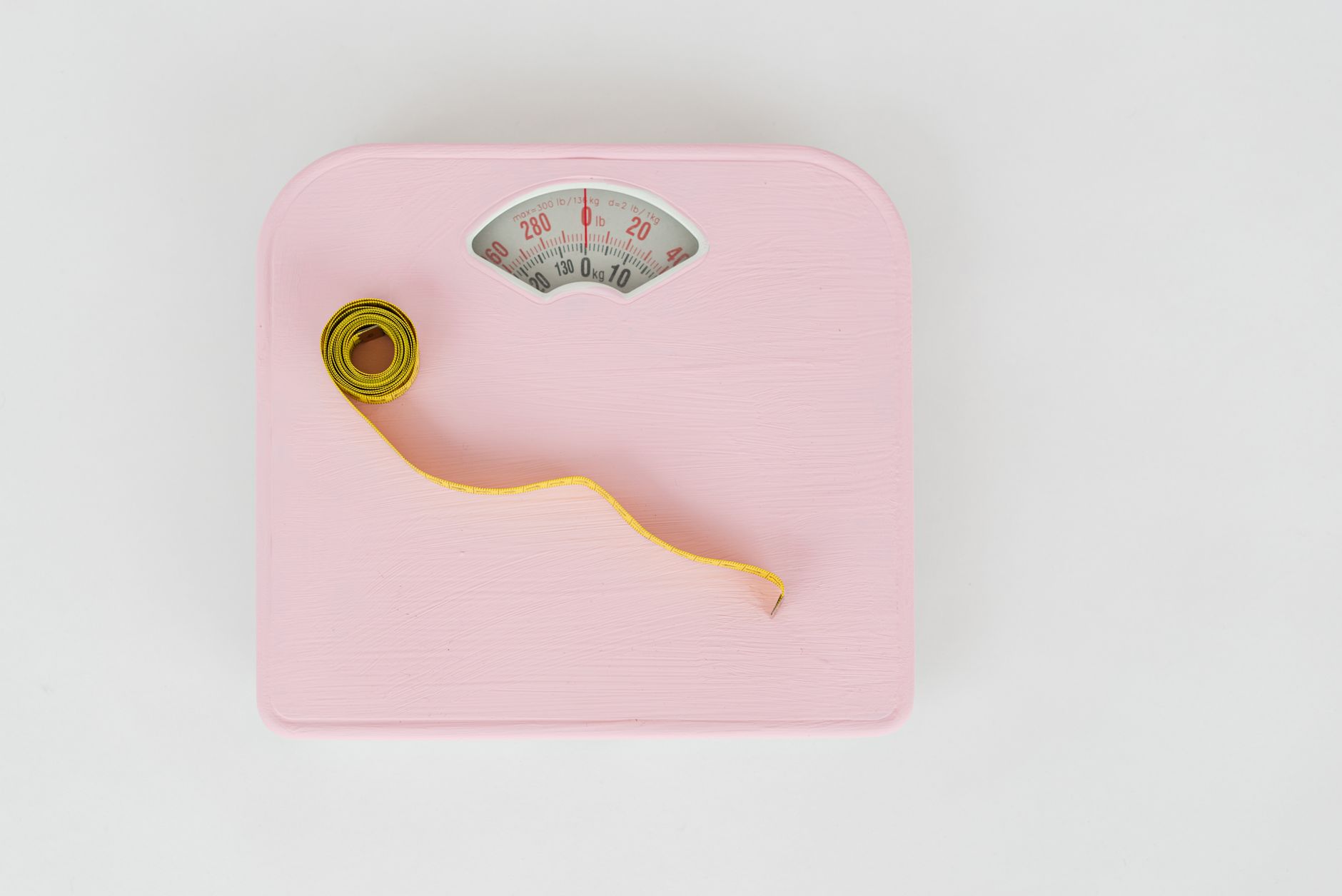
Set up a calm space and use a ruler or tape measure to gauge the gap.
What are the signs that my diastasis recti is getting worse after birth?
E
ven if your initial measurement was within the mild range, the separation can expand if the core is repeatedly overloaded. Watch for these warning signs:
Increasing width of the gap when you perform the self‑check.
Visible bulging or “pouching” in the midline when you lift, cough, or sneeze.
New or worsening lower‑back pain that feels deep rather than muscular soreness.
Difficulty engaging the “deep core” (the inner abdominal wall) during everyday tasks.
Persistent abdominal heaviness or a sense that the belly is “floppy.”
If any of these symptoms appear, especially bulging or pain, it’s time to schedule a professional evaluation. According to the American College of Obstetricians and Gynecologists (ACOG), progressive widening after the first six weeks postpartum warrants a targeted therapy plan.
Beyond the physical signs, pay attention to functional cues: trouble lifting a baby, difficulty standing from a seated position, or feeling that you “lose your balance” when you engage your core may all indicate that your diastasis is affecting daily life. Early detection lets you adjust activity levels before compensatory patterns become ingrained.
When should I see a physical therapist for diastasis recti after pregnancy?
Physical therapists (PTs) specializing in women's health are trained to assess the size of the separation, the quality of muscle recruitment, and the relationship to pelvic‑floor function. Consider seeing a PT if:
You have a gap larger than 4 cm on your self‑check.
You notice bulging, pain, or functional limitations.
You’re planning to return to exercise, sport, or heavy lifting and want a safe progression.
You’ve had a C‑section and are unsure how to protect the incision while strengthening the core.
Most experts recommend an initial PT visit between 6 and 12 weeks postpartum, once the uterus has involuted and you’re cleared for gentle movement by your OB‑GYN. Early referral can prevent maladaptive patterns that become harder to correct later.
During the first visit, the therapist will typically perform a standardized “finger‑width” measurement, assess your breathing pattern, and evaluate how well you can activate the transverse abdominis without bulging. This comprehensive picture guides a personalized program that balances core work, pelvic‑floor training, and safe progression of daily activities.
Can I do a diastasis recti check during my 6‑week postpartum visit?
Yes. The 6‑week postpartum appointment is a natural touchpoint for a quick diastasis assessment. Your OB‑GYN or midwife can perform a brief finger‑width test and discuss any concerns. However, the clinical exam may be brief, so you might still want to do a more detailed self‑check at home.
When you’re in the exam room, ask your provider:
“Can you show me how you’re measuring my diastasis?”
“What size of separation is considered normal for me?”
“Should I start core exercises now, or wait until I’ve re‑checked?”
Having a written record of your home measurement (date and gap size) can help your provider see trends over time.
Some clinics also offer a bedside ultrasound for a more precise picture, especially if the physical exam is inconclusive. Ultrasound can differentiate between a true diastasis and a small ventral hernia, which may require different management.
Difference between diastasis recti and a hernia in postpartum women
Both conditions involve a bulge in the abdominal wall, but they have distinct causes and treatment pathways. The table below highlights the key differences:
Feature
Diastasis Recti
Abdominal (Ventral) Hernia
Underlying Issue
Separation of the rectus abdominis muscles along the linea alba
Defect in the fascial layer allowing tissue or organ protrusion
Typical Gap Size
2–4 cm mild; > 4 cm moderate‑severe
Often > 2 cm but localized, may feel hard or tender
Pain Pattern
Diffuse, often low‑back or abdominal heaviness
Sharp, localized pain especially when straining
Risk Factors
Multiple pregnancies, large baby, rapid weight gain
Previous surgical incision, chronic coughing, heavy lifting
Treatment
Targeted core and pelvic‑floor exercises, PT, occasional surgery
Usually surgical repair (herniorrhaphy) if symptomatic
If you feel a firm, rope‑like bulge that appears only when you cough or lift, and especially if it’s painful, you may have a hernia rather than diastasis. An in‑person exam (or ultrasound) is the safest way to differentiate.
Because hernias can progress and risk incarceration, prompt medical evaluation is advised. In contrast, diastasis recti typically improves with conservative management and rarely requires urgent surgery.
How many centimeters apart is a normal postpartum diastasis recti measurement, and what are the measurement guidelines?
Guidelines from ACOG and the UK's National Institute for Health and Care Excellence (NICE) suggest the following ranges:
Normal: < 2 cm (roughly one finger‑width).
Mild: 2–4 cm (two to four finger‑widths).
Moderate: 4–5 cm (four to five finger‑widths).
Severe: > 5 cm (more than five finger‑widths).
These thresholds are based on research that links larger gaps with reduced trunk strength and higher rates of low‑back pain. Your own comfort and functional ability also matter—some women with a 3 cm gap feel perfectly fine, while others with a 2 cm gap notice weakness.
For consistency, always measure at the level of the umbilicus (belly button) and repeat the test after a short rest. Document the date, your position (lying or standing), and any symptoms you felt. This systematic record becomes a useful conversation starter with your provider.
Typical measurement ranges help you decide if your gap is mild, moderate, or severe.
Best exercises to improve diastasis recti after the initial check and other core work
Once you’ve confirmed the size of the separation, the goal is to re‑educate the deep core (the transverse abdominis) while protecting the linea alba. The following exercises are widely endorsed by ACOG, the NHS, and the International Association of Physical Therapists (IAPPT):
Pelvic‑tilt (supine march). Lie on your back, knees bent, and gently press the lower back into the floor while breathing out. This activates the deep core without creating bulge.
Heel slides. Slide one heel away while keeping the pelvis stable; engage the belly button toward the spine.
Modified side‑plank (knees down). Keep the body in a straight line, resting on the forearm and knees; hold for 10–15 seconds.
Standing pelvic floor contraction. While gently drawing the belly inward, contract the pelvic floor as if stopping a stream of urine. This dual activation supports the separation.
Diaphragmatic breathing with core engagement. Inhale deeply, allowing the belly to expand, then exhale while pulling the navel toward the spine.
Progress to more challenging moves—such as full side‑planks, bird‑dog, and gentle dead‑bugs—only after you can maintain a flat abdomen during the basic exercises. Avoid traditional crunches, sit‑ups, and heavy lifting until your gap is < 2 cm or your therapist advises otherwise.
Consistency matters more than intensity. Aim for short, focused sessions (5–10 minutes) several times a day rather than long, strenuous workouts that risk re‑opening the gap. Pair these moves with mindful breathing to keep intra‑abdominal pressure low.
How long does it take for diastasis recti to heal after delivery, including after C‑section?
Recovery timelines vary, but most research indicates:
Vaginal birth: 70 % of women see a reduction to < 2 cm by 12 weeks with consistent core work.
C‑section: Healing may be slower because the abdominal wall has an incision; many women reach similar results by 6‑month postpartum with guided PT.
Severe separations (> 5 cm): May require surgical repair if the gap does not improve after 6 months of dedicated therapy.
Key factors influencing speed are the size of the initial gap, adherence to exercise, nutrition (adequate protein and vitamin C), and avoidance of activities that increase intra‑abdominal pressure (heavy lifting, high‑impact cardio). The NHS notes that “most women regain functional core strength within 6 months, but full cosmetic resolution can take longer.”
It’s also normal to experience fluctuations; a slight increase in the gap after a bout of coughing or a new baby lift does not automatically signal failure. Track trends over weeks rather than day‑to‑day changes, and celebrate incremental progress.
Postpartum diastasis recti and pelvic floor connection
The abdominal and pelvic‑floor muscles work together like a corset. When the linea alba is widened, the pelvic floor often compensates by over‑activating, which can lead to urinary urgency or pelvic‑organ prolapse. Conversely, a weak pelvic floor can make the abdominal gap feel larger.
Integrating pelvic‑floor exercises—such as gentle Kegels performed while maintaining a drawn‑in abdomen—helps restore balance. A study from the International Urogynecological Association (IUGA) found that combined core‑pelvic‑floor programs improve both diastasis size and bladder control more effectively than core work alone.
Because the pelvic floor also stabilizes the lower spine, strengthening it can alleviate the low‑back discomfort that often accompanies diastasis. Think of the core as a “four‑wall box”: the front wall (abdominal muscles), the back wall (spine), and the top and bottom walls (diaphragm and pelvic floor). All four need to be engaged for optimal support.
Best postpartum core exercises for diastasis recti
For quick reference, here’s a weekly routine you can start after your 6‑week check, assuming a mild to moderate gap:
Saturday: Light yoga or stretching focusing on the thoracic spine and hips.
Sunday: Rest and body awareness—check your gap again if you feel any change.
Increase hold times or add a third set as you feel stronger, but keep the abdomen flat throughout each move. If you notice bulging, scale back and consult your therapist.
Remember to breathe naturally; holding your breath during these exercises can increase intra‑abdominal pressure and counteract your progress. A steady exhale while gently pulling the navel toward the spine is the safest cue.
How does breastfeeding affect diastasis recti healing?
Breastfeeding itself does not worsen diastasis, but the hormonal changes and the physical act of nursing can influence core tone. Oxytocin released during feeding promotes uterine involution, which can help the abdominal wall return to its pre‑pregnancy state. However, frequent lifting of the infant and the postural demands of nursing (often hunched over a baby) may place additional strain on the already‑stretched linea alba.
To support healing while nursing, keep your shoulders relaxed and use a supportive nursing pillow that brings the baby up to eye level, reducing forward‑head posture. Incorporate the same deep‑core exercises during feeding breaks—tiny pelvic‑tilts or diaphragmatic breaths can be done while holding your baby. If you notice increased bulging or pain while nursing, pause the activity and seek a PT assessment.
Hydration and adequate calories are also essential; lactation increases energy needs, and proper nutrition fuels tissue repair. Aim for a balanced diet rich in lean protein, leafy greens, and omega‑3 fatty acids, which have been linked to better muscle recovery.
Can abdominal binders help with diastasis recti?
Abdominal binders (often called postpartum belts) provide external support that can remind you to keep the core gently engaged, especially during daily activities like lifting a diaper bag or getting out of bed. ACOG notes that a well‑fitted binder may reduce discomfort and improve posture in the early weeks postpartum.
However, binders are not a substitute for active core training. Over‑reliance can lead to muscle “de‑conditioning,” where the deep core becomes weaker because it isn’t being used. If you choose to wear a binder, select one that offers gentle compression (not a tight corset) and wear it for short periods—typically 2–3 hours at a time—while you’re doing light chores.
Always discuss binder use with your OB‑GYN or PT, especially if you had a C‑section. A poorly placed binder can stress the incision site, potentially delaying healing.
When is surgery recommended for diastasis recti?
Most women improve with conservative care, but surgery becomes an option when the separation remains large (> 5 cm) after 6–12 months of dedicated therapy, or when the diastasis causes persistent pain, functional limitation, or significant cosmetic concerns that affect quality of life.
Procedures typically involve “abdominoplasty with plication,” where the surgeon sutures the separated rectus muscles back together. The American Society of Plastic Surgeons (ASPS) recommends that surgery be performed by a board‑certified surgeon with experience in postpartum abdominal reconstruction.
Pre‑operative evaluation includes a thorough physical exam, imaging (often ultrasound), and a discussion of risks such as infection, seroma, or recurrence. Post‑operative care mirrors that of a C‑section: limited lifting, use of a binder, and a gradual return to core exercises under PT supervision.
Because surgery carries inherent risks, it’s considered a last‑resort option. Most women find that continued PT, proper nutrition, and patience lead to satisfactory functional and aesthetic outcomes without the need for an operation.
From our medical team: “A postpartum diastasis recti check is a simple, safe way to monitor your core healing. Most women improve with gentle, consistent exercise and by avoiding high‑impact activities early on. If the gap isn’t shrinking or you develop pain, we recommend a professional assessment to rule out a hernia or to discuss surgical options.”
Myth vs. fact
Myth: “If I can’t see a gap, I don’t have diastasis recti.”
Fact: The separation can be hidden by body fat or muscle tone. A proper finger‑width measurement is the most reliable method.
Myth: “Crunches will close the gap faster.”
Fact: Traditional crunches increase intra‑abdominal pressure and can worsen the separation. Targeted deep‑core work is the evidence‑based approach.
Myth: “Only women who have a C‑section get diastasis recti.”
Fact: Pregnancy itself stretches the linea alba, so both vaginal and C‑section deliveries can lead to diastasis.
Key takeaways
Measure the gap at the belly button with a ruler or soft tape; < 2 cm is normal.
Perform the self‑check lying flat, using a gentle head‑lift to feel the separation.
Red‑flag signs include bulging, increasing width, and new lower‑back pain.
Seek a physical therapist if the gap exceeds 4 cm or you notice functional limits.
Gentle core exercises (pelvic‑tilt, heel slides, modified side‑plank) support healing.
Most mild cases improve within 12 weeks; severe separations may need surgery.
Breastfeeding does not hinder recovery, but maintain good posture and stay hydrated.
Abdominal binders can provide temporary support but should not replace active core work.
Frequently asked questions
How can I tell if I have diastasis recti after giving birth?
Start with a simple finger‑width test: lie on your back, gently lift your head, and measure the distance between the two sides of your abdominal muscles at the belly button. A gap of < 2 cm is normal; larger gaps indicate diastasis.
Is it safe to do crunches with diastasis recti?
Generally no—crunches increase intra‑abdominal pressure and can widen the gap. Instead, focus on deep‑core moves like pelvic‑tilts and diaphragmatic breathing until your gap shrinks.
When will my diastasis recti heal on its own?
Most mild separations (< 4 cm) improve naturally within 3–4 months with gentle core work. Larger gaps may take 6 months or longer, and some may need surgical repair if they don’t reduce after consistent therapy.
What is the normal width of a postpartum diastasis recti?
Guidelines from ACOG and NICE define a normal width as less than 2 cm (about one finger‑width). Anything above that is considered a mild to severe separation depending on the exact measurement.
Can diastasis recti cause lower back pain?
Yes. When the abdominal wall is weakened, the lower back muscles often compensate, leading to strain and discomfort. Strengthening the deep core and pelvic floor can alleviate this pain.
Should I see a doctor for a postpartum diastasis recti check?
If you notice a gap larger than 2 cm, experience bulging, or have pain, it’s wise to schedule a visit with your OB‑GYN or a women’s‑health physical therapist. Even mild cases benefit from professional guidance to ensure safe exercise progression.
Can I continue running or high‑impact cardio with diastasis recti?
Low‑impact cardio (walking, elliptical, swimming) is generally safe once you can keep the abdomen flat during the activity. High‑impact exercises that cause jolting or heavy landing should be paused until your gap is < 2 cm or a therapist clears you.
How often should I re‑measure my diastasis recti?
Re‑measure every 2–4 weeks in the first three months postpartum, then monthly until the gap stabilizes. Consistent tracking helps you spot trends and adjust your exercise plan with your therapist.
When to call your doctor
If you experience any of the following, contact your provider promptly:
Sudden increase in the size of the abdominal bulge.
Sharp, localized pain that worsens with coughing or lifting.
Visible protrusion that feels hard or tender (possible hernia).
New urinary incontinence or pelvic‑organ pressure.
Fever, redness, or drainage from a C‑section incision combined with abdominal changes.
This information is for educational purposes only and does not replace professional medical advice.
References
American College of Obstetricians and Gynecologists. “Physical Activity and Exercise During Pregnancy and the Postpartum Period.” ACOG Practice Bulletin No. 229, 2023.
National Institute for Health and Care Excellence (NICE). “Postnatal Care: Routine Checks and Interventions.” NICE Guideline NG123, 2022.
National Health Service (NHS). “Diastasis Recti – Symptoms and Treatment.” NHS.uk, updated 2023.
International Association of Physical Therapists (IAPPT). “Core Rehabilitation After Pregnancy.” Journal of Women’s Health Physical Therapy, 2021.
International Urogynecological Association (IUGA). “Combined Core and Pelvic Floor Rehabilitation for Postpartum Diastasis Recti.” IUGA Clinical Review, 2022.
Mayo Clinic. “Diastasis Recti.” Mayo Clinic Proceedings, 2022.
World Health Organization (WHO). “Maternal Health Guidelines – Postpartum Care.” WHO Publication, 2021.
Centers for Disease Control and Prevention (CDC). “Postpartum Care Overview.” CDC.gov, 2023.
La Leche League International. “Breastfeeding and Postpartum Recovery.” LLLI Guidance, 2022.
American Society of Plastic Surgeons (ASPS). “Abdominoplasty for Diastasis Recti.” ASPS Patient Education, 2023.
Not sure about the label on Postpartum Diastasis Recti Check products?
Snap the ingredients list and SafeFilter checks every ingredient for your stage — only 3 free scans this month, then you're locked until reset. Unlimited from $7/mo or $50/yr.
When Shubhra Mishra was expecting her first child in 2016, she was overwhelmed by conflicting food advice — one site said yes, another said never. By the time her second baby arrived in 2019, she realized millions of mothers face the same confusion.
That sparked a five-year journey through clinical nutrition papers, cultural diets, and expert conversations — all leading to BumpBites: a calm, compassionate space where science meets everyday motherhood.
Her long-term vision is to build a global community ensuring safe, supported, and free deliveriesfor every mother — because no woman should face pregnancy alone or uninformed. 🌿
🌍 Stand with mothers, shape safer guidance
Join a small circle of experts who review BumpBites articles so expecting parents everywhere can decide with confidence.