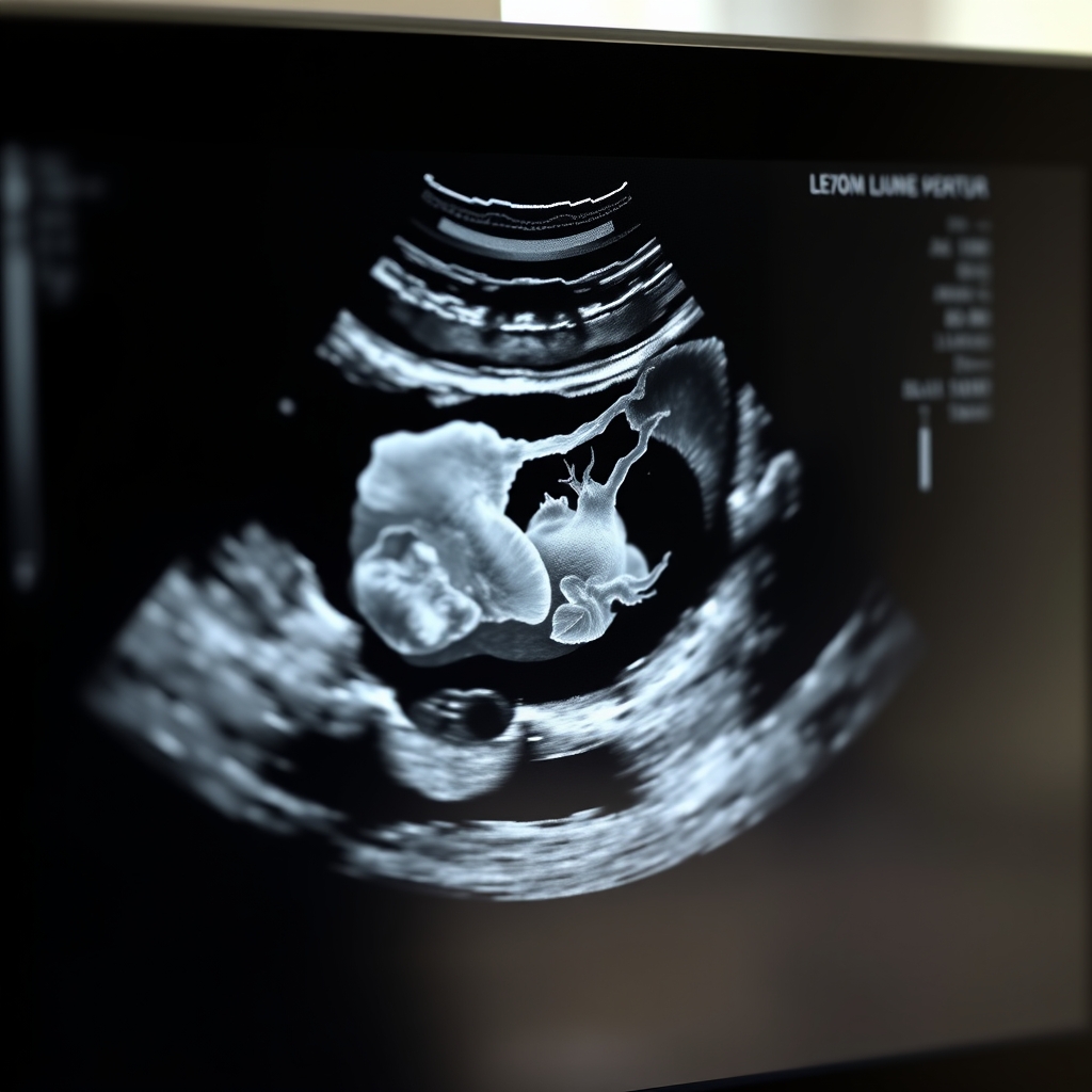
Find out when you can see your baby on a sonogram, the earliest sonogram for pregnancy is typically around 5-6 weeks, learn more
By Shubhra Mishra — a mom of two who turned her own confusion during pregnancy into BumpBites, a global mission to make food choices clear, safe, and stress-free for every expecting mother. 💛
Check whether any food is safe during pregnancy with the BumpBites Food Safety Checker.
Quick take: You can usually see a gestational sac as early as 5 weeks, a heartbeat around 6 weeks, and a clearer embryo by 7 weeks. Transvaginal scans are the most sensitive early on, and both ACOG and NHS say early ultrasounds are safe when medically indicated.
It’s 2 a.m., you’ve just taken a pregnancy test, and the excitement is tangled with a pinch of anxiety. “Can I already see the baby?” you wonder, scrolling through endless articles that promise a glimpse of the tiny heart beating inside. You’re not alone—many expectant parents wonder exactly when the first sonogram will reveal something beyond a blurry circle.
In this guide we’ll walk through the timeline of the earliest ultrasound views, explain the differences between transvaginal and abdominal scans, and show you what structures you can expect at 5, 6, and 7 weeks. We’ll also cover safety, accuracy, and how doctors use these early images to date the pregnancy and spot multiples. By the end you’ll know exactly when you can expect to see your baby on a scan, what to look for, and which questions to ask your provider.
Whether you’re planning your first appointment or just curious about the “baby‑watch” milestones, we’ve gathered the most up‑to‑date guidance from ACOG, the NHS, and other leading bodies. Let’s start with the moment you’ll first catch a glimpse of your growing little one.
When can I first see my baby on a sonogram?
The very first sonographic sign of pregnancy is the gestational sac, a fluid‑filled pocket that houses the embryo. Most clinicians can spot this sac at 5 weeks gestation (counting from the first day of your last menstrual period). At this stage the image looks like a small, round bubble within the uterus, and it’s often the only thing visible.
By the end of week 5, the sac usually measures around 6–8 mm. A faint yolk sac may appear alongside it, providing the embryo’s first source of nutrition. However, the embryo itself is still too tiny to be distinguished on an abdominal scan, and a heartbeat is usually not yet detectable.
Most providers will schedule a follow‑up scan at 6–7 weeks to confirm that the embryo is developing as expected. If you’re eager to see a tiny flicker of movement, a transvaginal scan can often capture a heartbeat as early as 5 weeks + 4 days. The key takeaway: the first “baby” you’ll see is the sac, not the little person, and it appears around the five‑week mark.
Why the five‑week window matters: At this point the uterus is still small enough to sit deep in the pelvis, which is why a transvaginal probe—placed just a few centimeters from the gestational sac—provides the clearest view. If you’re a first‑time mom, seeing that tiny sac can be an emotional reassurance that the pregnancy is progressing, even before you feel any fetal movements.
At about 5 weeks, the gestational sac is the first structure you’ll see on a scan.
What week does the first ultrasound show the heartbeat?
The first reliable detection of a fetal heartbeat typically occurs at 6 weeks gestation. In a transvaginal scan, the cardiac activity can be seen when the embryo reaches a crown‑rump length (CRL) of roughly 5–6 mm. The heartbeat usually pulses at 90–110 beats per minute (bpm) at this stage, and it speeds up rapidly over the next weeks.
On an abdominal scan, the heartbeat often becomes visible a little later—around 6 weeks + 2 days—because the probe is farther from the uterus and the image resolution is lower. If you have a particularly early scan and the heart isn’t seen yet, your provider may simply recommend a repeat in a few days.
It’s worth noting that a “missing heartbeat” does not automatically mean a problem. Embryonic development can vary by a few days, and occasional false‑negative results happen, especially with abdominal imaging. Always follow up with your clinician if you’re unsure.
What the heartbeat tells you: Beyond confirming viability, the heart rate is a useful marker of embryonic health. A slower rate (below 90 bpm) at 6 weeks may signal growth restriction, while an unusually fast rate can be an early sign of certain chromosomal conditions. Your sonographer will note the exact beats per minute, and your provider will interpret it in the context of other findings.
Earliest safe time for a pregnancy ultrasound
Both the American College of Obstetricians and Gynecologists (ACOG) and the UK’s National Health Service (NHS) state that ultrasound is safe at any stage when medically indicated. The technology uses non‑ionizing sound waves, which have not been shown to cause harm to the fetus or mother when used appropriately.
Safety guidelines focus on indication rather than gestational age. In other words, an ultrasound is considered safe if there’s a clear clinical reason—such as confirming a viable pregnancy, dating the pregnancy, or checking for multiples. Routine “keeps‑ake‑the‑baby‑happy” scans without a medical purpose are discouraged, primarily to avoid unnecessary expense and potential over‑diagnosis.
For early scans, the ALARA principle (“as low as reasonably achievable”) guides sonographers to use the lowest possible acoustic output while still obtaining a diagnostic image. This principle is built into the training standards of both ACOG and the Royal College of Obstetricians and Gynaecologists (RCOG).
What “safe” means in practice: The FDA’s 2023 safety communication emphasizes that typical obstetric ultrasound exposure is far below the threshold for any known biological effect. In the United Kingdom, the NHS’s “Diagnostic Imaging” policy requires that each scan be justified and documented, ensuring that no unnecessary exposure occurs. If you have concerns about cumulative scans, ask your provider to review the total number and timing of any previous imaging.
How accurate is a 6 week sonogram for detecting pregnancy?
A 6‑week transvaginal sonogram is highly accurate for confirming an intrauterine pregnancy. Studies cited by ACOG show that a gestational sac at this stage predicts a live birth in roughly 80–85 % of cases. The presence of a yolk sac and a detectable heartbeat further increase the likelihood of a healthy pregnancy.
However, accuracy for dating the pregnancy improves after 8 weeks, when fetal measurements become more consistent. At 6 weeks, the CRL can vary by up to 7 days, which translates into a broader range for estimated due date (EDD). Consequently, many providers use the 6‑week scan primarily to confirm viability and then schedule a 10‑ to 12‑week scan for precise dating.
Detecting multiples (twins or higher-order) at 6 weeks is also possible, though the embryos may be too close together to differentiate clearly. If twins are suspected, a follow‑up scan at 8–10 weeks typically provides a definitive picture.
Limitations to keep in mind: A 6‑week scan may miss a very early pregnancy loss that occurs after the scan but before the next appointment. For this reason, many clinicians advise a “watchful waiting” period of a few days to a week before drawing conclusions about viability if the early scan is ambiguous.
What does a 7 week ultrasound image look like?
By the end of week 7, the embryo grows to a CRL of about 7‑9 mm. On a transvaginal scan you’ll see a more defined shape with the beginnings of limb buds, a distinct head, and a clearly visible beating heart. The heartbeat usually ranges between 140–170 bpm, a noticeable acceleration from week 6.
The yolk sac remains prominent, and you may also spot the amniotic fluid pocket surrounding the embryo. The image often resembles a tiny bean or a “flying saucer” with a dark center (the embryo) and a brighter ring (the yolk sac). This is the first stage where you can truly say you’re seeing a “baby” rather than just a sac.
On an abdominal scan, the 7‑week image may still be slightly less detailed, but most sonographers can capture the heart and basic limb structures. If you’re hoping for a clear picture, a transvaginal approach remains the gold standard for this early window.
What to expect emotionally: Many parents describe a mix of awe and relief when they first see the flickering heart. It’s a concrete sign that the pregnancy is progressing, and it often eases anxiety that can build during the first trimester. If you’re unable to attend the scan in person, many clinics now offer secure digital images so you can share the moment with partners or family members.
At 7 weeks, the embryo’s heartbeat and early limb buds become visible.
Differences between transvaginal and abdominal early sonograms
Both transvaginal (TV) and abdominal (AB) ultrasounds use the same sound‑wave technology, but the probe placement and image quality differ markedly in early pregnancy.
Feature
Transvaginal (TV)
Abdominal (AB)
Earliest detectable structure
Gestational sac at 5 weeks (≈6 mm)
Gestational sac at 6 weeks (≈8 mm)
Heartbeat detection
5 weeks + 4 days
6 weeks + 2 days
Image clarity
High resolution, detailed embryo
Lower resolution, less detail
Comfort
Invasive, requires a speculum
Non‑invasive, fully external
Typical cost (US)
$150–$200
$100–$150
Best for
Early dating, detecting heartbeat, evaluating viability
Later first‑trimester scans, routine check‑ups
Because the TV probe sits just a few centimeters from the uterus, it can capture finer details earlier, making it the preferred method for confirming a pregnancy before 6 weeks. The AB approach is more comfortable for many patients and is sufficient once the fetus grows beyond 8 weeks.
Both methods adhere to the same safety standards; the only difference is the level of acoustic output, which is kept low for both types of scans. If you have a history of pelvic pain or a strong aversion to internal exams, discuss the possibility of an abdominal scan with your provider—they may still obtain useful images, just a bit later.
Choosing the right scan for you: Some clinics offer a “combined” approach—starting with a transvaginal scan to locate the sac quickly, then switching to an abdominal scan for a broader view. This can balance comfort with diagnostic detail, especially for patients who are nervous about internal probes.
What are the signs of a viable pregnancy on early ultrasound?
Viability on early ultrasound is judged by a combination of visual landmarks:
Gestational sac – a well‑defined, round structure within the uterine cavity.
Yolk sac – appears as a small, circular echo inside the sac; its presence supports early development.
Embryo – a tiny, darker object within the sac, usually visible by 6 weeks.
Cardiac activity – a rhythmic flicker indicating a heartbeat; the hallmark of viability.
Mean sac diameter (MSD) – measurements that correlate with gestational age; an MSD of 10–12 mm at 5 weeks is typical.
At 6 weeks, a heartbeat of 90–110 bpm is reassuring. By 7 weeks, the heart rate climbs to 140–170 bpm, and limb buds become discernible. If any of these features are missing, your provider may repeat the scan in a few days to see if the embryo is simply developing later.
Other reassuring signs include a consistent crown‑rump length (CRL) measurement that falls within the expected range for the gestational age, and a yolk sac size of 2–5 mm. Larger yolk sacs or irregular shapes can sometimes signal a non‑viable pregnancy, but only a trained sonographer can interpret those nuances.
When things look atypical: A “blighted ovum” (an empty gestational sac) may appear as a sac without an embryo after 6 weeks. In such cases, clinicians usually schedule a follow‑up scan in 7–10 days to confirm whether development proceeds. This cautious approach respects both the emotional impact on parents and the biological variability of early embryos.
Key structures to look for on a 5‑ to 7‑week scan.
Earliest ultrasound to confirm due date and how many weeks until baby is visible on ultrasound
The most accurate dating scan is typically performed between 8 and 12 weeks. At this point the fetal crown‑rump length (CRL) correlates strongly with gestational age, giving an estimated due date (EDD) that is within ±5 days of the actual date. Earlier scans (5‑7 weeks) can confirm pregnancy but are less precise for dating because the embryo’s growth rate varies more in the first few weeks.
If you’re eager to know the exact date, ask your provider about a “dating scan” at 10 weeks. This scan will also assess the placenta’s location and check for any early anomalies. While a 5‑week scan can tell you “yes, we see a sac,” it cannot reliably predict the due date with the same confidence as a later scan.
In terms of visibility, the baby (embryo) becomes “clearly visible” on most scans by 7 weeks. Before that, you’re mainly seeing the sac and yolk sac. The timeline looks like this:
5 weeks – Gestational sac, possibly yolk sac.
5 weeks + 4 days – Possible heartbeat on transvaginal.
6 weeks – Embryo visible, heartbeat usually present.
8 weeks – Better definition, early organ formation.
While a 5‑week scan can sometimes detect twins (two separate sacs), confirming multiple gestations is more reliable at 7 weeks or later when each embryo has its own heartbeat and distinct sac.
Practical tip: If you’re planning a vacation or have a busy work schedule, aim for the 8‑ to 10‑week dating scan. That window gives you the most accurate due date while still allowing enough time to make any necessary adjustments to prenatal care or travel plans.
How to prepare for your early ultrasound appointment
Preparing for a first‑trimester scan is simple, but a few small steps can improve image quality and make the experience smoother. For a transvaginal scan, you’ll be asked to empty your bladder beforehand; a full bladder can actually make the probe harder to maneuver. For an abdominal scan, a comfortably full bladder (about 1 cup of water) helps lift the uterus into a better position for imaging.
Wear loose‑fitting clothing that can be easily pulled up or down, and bring a list of any medications you’re taking, including prenatal vitamins. If you have a history of pelvic surgery, scar tissue can affect image clarity, so let the sonographer know ahead of time. Many clinics also allow you to bring a partner or support person; having a familiar face in the room can reduce anxiety.
What to expect during the exam: The sonographer will apply a warm, water‑based gel to the skin (or a speculum for a transvaginal scan) to ensure good contact with the probe. The procedure itself usually lasts 10–20 minutes. You’ll hear the gentle hum of the machine and may see a monitor displaying the real‑time image. Feel free to ask the technician to point out structures you’re curious about—their explanations can turn a technical image into a memorable moment.
What to do if the early scan is inconclusive or shows no heartbeat
It’s rare, but sometimes an early scan doesn’t reveal a heartbeat or a clear embryo. The most common reasons are that the pregnancy is simply a few days younger than expected, or that the embryo is positioned in a way that makes visualization difficult. In these cases, clinicians usually recommend a repeat scan in 5–7 days rather than jumping to conclusions.
If a repeat scan still shows no cardiac activity, your provider may discuss the possibility of a miscarriage and offer options such as expectant management, medical treatment, or surgical intervention, depending on your preferences and clinical situation. Throughout this process, emotional support is essential—consider reaching out to a counselor, support group, or trusted friend.
Key reassurance: Even when an early scan is ambiguous, most pregnancies continue to develop normally. The timing of the first heartbeat can vary, and many parents report that the “first picture” of their baby comes a little later than they hoped. Patience and open communication with your care team are the best strategies.
Doctor’s note
From our medical team: Early ultrasound is a valuable tool for confirming a viable intrauterine pregnancy and establishing an accurate timeline. The most common concerns—radiation exposure and fetal harm—are unfounded when the scan follows professional guidelines. If you have a history of miscarriage, pre‑eclampsia, or known uterine anomalies, your provider may recommend an earlier transvaginal scan to monitor development closely. Always discuss any anxiety you feel about the procedure; a calm environment and a skilled sonographer can make the experience both reassuring and informative.
Myth vs. fact
Myth: “Early ultrasounds can damage the baby’s brain.”
Fact: Ultrasound uses sound waves, not ionizing radiation, and extensive research by ACOG and the FDA shows no evidence of harm when performed by trained professionals.
Myth: “If I don’t see a heartbeat at 5 weeks, something is wrong.”
Fact: The heartbeat may not be detectable until 6 weeks, especially on an abdominal scan. A repeat scan in a few days is the standard approach.
Myth: “You can see the baby’s face at 6 weeks.”
Fact: At 6 weeks the embryo is only a tiny bean‑shaped structure; facial features develop later, typically after 8 weeks.
Key takeaways
The gestational sac appears around 5 weeks; the heartbeat usually shows up at 6 weeks.
Transvaginal scans detect structures earlier and are the preferred method for confirming viability before 7 weeks.
Early ultrasounds are safe when performed for a medical indication; there’s no proven risk to the fetus.
Accurate dating is best done between 8‑12 weeks, when crown‑rump length measurements are most reliable.
If you’re unsure about what you saw, schedule a repeat scan—development can vary by a few days.
Ask your provider about the scan’s purpose, preparation steps, and what you’ll see on the images.
Stay calm, bring a support person, and remember that many early scans are just the first glimpse of a journey that will become clearer each week.
Frequently asked questions
When can you first see the baby on an ultrasound?
The first visual cue is the gestational sac at about 5 weeks; a clear embryo and heartbeat are usually visible by 6–7 weeks.
Is a 6‑week ultrasound safe for the baby?
Yes. Both ACOG and the NHS consider early ultrasound safe when medically indicated; the sound waves do not harm fetal tissue.
What does a 7‑week pregnancy ultrasound show?
At 7 weeks you’ll typically see a tiny embryo with a beating heart, early limb buds, and a prominent yolk sac.
Can a transvaginal ultrasound detect a heartbeat earlier than an abdominal one?
Exactly. A transvaginal probe can pick up cardiac activity as early as 5 weeks + 4 days, while an abdominal scan usually sees it around 6 weeks + 2 days.
How accurate is an early sonogram in dating pregnancy?
Dating is most accurate after 8 weeks; a 6‑week scan can confirm viability but may be off by up to a week in estimating the due date.
What are the signs of a healthy pregnancy on the first ultrasound?
Key signs include a well‑defined gestational sac, a visible yolk sac, an embryo with a measurable crown‑rump length, and a rhythmic heartbeat.
Do I need to fast before a first‑trimester ultrasound?
No fasting is required. For an abdominal scan, a comfortably full bladder (about one cup of water) helps improve image quality, while a transvaginal scan is performed with an empty bladder.
Can I have an ultrasound if I have a pacemaker or other implanted device?
Yes. Ultrasound does not use electromagnetic fields that interfere with pacemakers, so it is generally safe. However, always inform the sonographer of any implants so they can document it in the report.
When to call your doctor
If you experience any of the following, contact your obstetrician or midwife right away: severe abdominal pain, heavy bleeding, fever over 100.4 °F (38 °C), sudden loss of pregnancy symptoms, or if you’re unable to locate a heartbeat on a scheduled scan. This article is for informational purposes only and does not replace personalized medical advice.
References
American College of Obstetricians and Gynecologists. “Ultrasound in Pregnancy.” ACOG Committee Opinion No. 730, 2021.
National Health Service (NHS). “Ultrasound scans in pregnancy.” Clinical guideline CG190, 2022.
Royal College of Obstetricians and Gynaecologists (RCOG). “Early Pregnancy Ultrasound.” Green‑top Guideline No. 70, 2020.
Food and Drug Administration (FDA). “Ultrasound Safety.” 2023 safety communication.
World Health Organization (WHO). “Recommendations for Antenatal Care.” 2022.
Society of Radiology. “Principles of ALARA for Diagnostic Imaging.” 2021.
Centers for Disease Control and Prevention (CDC). “Pregnancy Planning and Early Prenatal Care.” 2022.
American Pregnancy Association. “Early Ultrasound FAQ.” Updated 2023.
Not sure about the label on Earliest Sonogram For Pregnancy When You Can See Baby products?
Snap the ingredients list and SafeFilter checks every ingredient for your stage — only 3 free scans this month, then you're locked until reset. Unlimited from $7/mo or $50/yr.
When Shubhra Mishra was expecting her first child in 2016, she was overwhelmed by conflicting food advice — one site said yes, another said never. By the time her second baby arrived in 2019, she realized millions of mothers face the same confusion.
That sparked a five-year journey through clinical nutrition papers, cultural diets, and expert conversations — all leading to BumpBites: a calm, compassionate space where science meets everyday motherhood.
Her long-term vision is to build a global community ensuring safe, supported, and free deliveriesfor every mother — because no woman should face pregnancy alone or uninformed. 🌿
🌍 Stand with mothers, shape safer guidance
Join a small circle of experts who review BumpBites articles so expecting parents everywhere can decide with confidence.