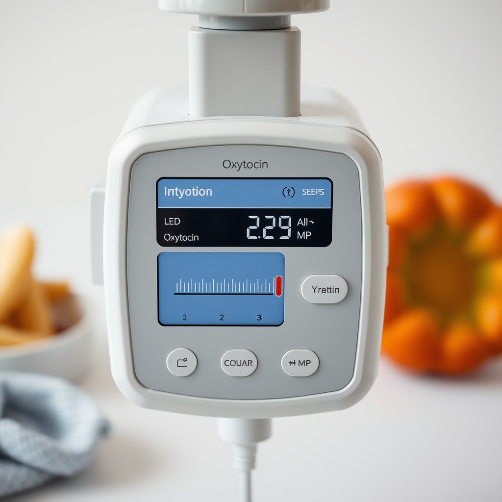
VBAC induction oxytocin titration + uterine rupture surveillance: learn how oxytocin is used to induce labor in VBAC with careful monitoring
By Shubhra Mishra — a mom of two who turned her own confusion during pregnancy into BumpBites, a global mission to make food choices clear, safe, and stress-free for every expecting mother. 💛
Check whether any food is safe during pregnancy with the BumpBites Food Safety Checker.
Quick take: Inducing a VBAC with oxytocin is possible when the mother meets strict eligibility criteria, the hospital follows a low‑dose titration protocol, and continuous maternal‑fetal surveillance is in place to catch any sign of uterine rupture quickly.
It’s 2 a.m., you’ve just finished a glass of water and are scrolling through “VBAC induction” because the clock is ticking and you’ve been told you’re a good candidate for a trial of labor after cesarean. The worry in the back of your mind is simple: “Will the baby come safely, or could my uterus give way?” You’re not alone—many expectant parents face the same question when a previous C‑section meets a scheduled induction.
🔢 Calculate it for your situation: Use our Oxytocin Titration for a personalized result in seconds.
Below you’ll find a step‑by‑step guide that walks you through the whole process—who can try it, how oxytocin is carefully titrated, what monitoring looks like, how clinicians spot a uterine rupture early, and what to expect compared with spontaneous labor. We also cover the staffing, equipment, and policy pieces that keep a VBAC induction safe, plus a quick calculator link for the exact infusion rates you’ll hear about in the delivery room.
Bottom line: When eligibility criteria are met, a low‑dose oxytocin protocol with vigilant surveillance can be as safe as a spontaneous VBAC, but it requires strict adherence to guidelines and rapid response plans.
Who is eligible for a VBAB induction, and what are the contraindications?
Eligibility hinges on both the mother’s obstetric history and the current pregnancy’s characteristics. The American College of Obstetricians and Gynecologists (ACOG) and the Royal College of Obstetricians and Gynaecologists (RCOG) list the same core factors, though exact wording can differ between the United States and the United Kingdom.
Core eligibility criteria
One prior low transverse cesarean scar (the most common and safest type).
Gestational age ≥ 37 weeks; most hospitals start induction at 39 weeks to reduce the need for early‑term delivery.
Singleton pregnancy with cephalic (head‑down) presentation.
No contraindication to vaginal birth such as placenta previa, active genital herpes, or severe pre‑eclampsia.
Maternal comorbidities that are well‑controlled (e.g., hypertension, diabetes) and do not demand immediate delivery.
Key contraindications for VBAC induction
Classical (vertical) uterine incision or T‑shaped scar—these carry a higher rupture risk.
Multiple prior cesarean sections, especially if any were non‑low‑transverse.
Uterine anomalies (e.g., bicornuate uterus) that compromise wall integrity.
Fetal malpresentation (breech or transverse) that would require a cesarean anyway.
Maternal conditions that mandate a rapid delivery (severe fetal distress, uncontrolled hemorrhage).
When a provider determines you meet the eligibility checklist, the next step is to decide whether induction is medically necessary (e.g., post‑dates pregnancy, pre‑eclampsia, or maternal request) or whether you can await spontaneous labor. Induction is not automatically a red flag, but it does require a more stringent monitoring plan.
In practice, many clinicians also assess cervical favorability (Bishop score) before deciding on the induction method. A score of 6 or higher usually predicts a smoother labor, while a lower score may prompt the use of mechanical ripening before oxytocin is started.
It’s worth noting that eligibility is not static; a woman who initially seems borderline may become a clear candidate after a short period of cervical ripening, which can improve the odds of a successful vaginal delivery without raising rupture risk.
Oxytocin titration protocol for VBAC induction
Oxyto
cin (Pitocin) is the most common medication for cervical ripening and labor augmentation. In a VBAC setting, the protocol starts at a lower dose and climbs more slowly than in a primary‑cesarean cohort to give the uterine scar time to adjust.
Step‑by‑step dosing schedule
Baseline assessment: Confirm a reassuring fetal heart rate (FHR) pattern, maternal vitals, and a fully empty bladder.
Start infusion: Begin with 0.5 mU/min (millunits per minute) of oxytocin diluted in 1000 mL of normal saline.
First titration point (20 minutes): If no uterine contractions (defined as ≥ 3 contractions in 10 minutes) and FHR remains normal, increase to 1.0 mU/min.
Second titration point (20 minutes later): If still inadequate, raise to 2.0 mU/min.
Maximum dose: For VBAC, many institutions cap the infusion at 10 mU/min, but many stop earlier (6 mU/min) if contractions become too strong (≥ 5 contractions/10 min) or if fetal tracing shows decelerations.
Pause and reassess: Once adequate labor (≥ 3 contractions/10 min with average intensity ≥ 30 mmHg) is achieved, the infusion is held steady for 2 hours, then weaned down by 0.5–1 mU/min every 30 minutes.
Because each hospital may have its own infusion pump settings, we recommend using the BumpBites Oxytocin Titration calculator to double‑check the exact numbers for your unit’s equipment.
Why a low‑dose approach matters
Studies from ACOG and NICE (National Institute for Health and Care Excellence) show that a low‑dose protocol halves the incidence of uterine rupture compared with a standard dose (0.2 mU/min increase every 15 minutes). The slower climb gives the scar tissue time to stretch gradually, reducing the shear forces that can create a rupture.
In addition, a gentler rise in uterine activity is associated with fewer episodes of hyperstimulation, which can otherwise lead to fetal distress and a cascade of interventions. This is why many hospitals have adopted the “low‑dose, high‑vigilance” model as their default for VBAC inductions.
Adjustments for special situations
Maternal obesity (BMI ≥ 30): May need a slightly higher ceiling (up to 12 mU/min) because subcutaneous fat can blunt drug absorption.
Pre‑existing uterine scar tenderness: If the mother reports localized pain, pause the infusion and assess with bedside ultrasound.
Fetal monitoring changes: Any late decelerations or persistent variable decelerations should prompt a dose reduction or temporary stop.
Accurate low‑dose titration is the cornerstone of a safe VBAC induction.
In addition to the standard titration, many centers now incorporate a “pause‑point” after the first 2 mU/min increase to allow the uterus to adapt before the next escalation. This practice, endorsed by the Society for Maternal‑Fetal Medicine (SMFM), can further reduce hyperstimulation and the associated risk of rupture.
When a patient’s labor does not progress after reaching the dose ceiling, clinicians may consider a brief “drug holiday” to let the uterus rest before resuming a slower titration, a strategy that has been described in recent ACOG guidance updates.
Continuous maternal and fetal monitoring during VBAC induction
Surveillance is the safety net that catches the rare but serious event of uterine rupture. Both maternal and fetal parameters are watched around the clock, often with the same equipment used for standard inductions, but with a higher threshold for alerts.
Maternal monitoring components
Uterine activity: External tocodynamometer or intrauterine pressure catheter (IUPC) if the external trace is unclear. Target is 3–5 contractions per 10 minutes, each lasting 40–60 seconds.
Blood pressure and heart rate: Checked every 15 minutes during the active phase, more frequently if the mother has hypertension.
Pain assessment: Sudden, sharp, or “tearing” pain, especially in the lower abdomen or back, is a red flag for rupture.
Maternal urine output: Maintained > 0.5 mL/kg/h; oliguria can signal early hemorrhage.
Fetal monitoring guidelines
Continuous electronic fetal monitoring (EFM): Recommended for all VBAC inductions, per ACOG Practice Bulletin 2020.
Baseline FHR: 110–160 bpm; variability should be moderate to marked.
Decelerations: Late decelerations or prolonged variable decelerations (> 60 seconds) trigger an immediate evaluation.
Biophysical profile (BPP) or scalp pH sampling: Considered if non‑reassuring patterns persist despite interventions.
Documentation and communication
Every change in contraction strength, maternal vitals, or fetal tracing is logged in real time. The bedside nurse alerts the laborist or obstetrician the moment a threshold is crossed, and a second nurse double‑checks the reading. This redundancy is essential in low‑resource settings where staffing may be thin; a dedicated “VBAC induction” champion on each shift can help maintain vigilance.
Electronic medical records (EMR) often include a “VBAC induction” order set that auto‑populates monitoring intervals, ensuring no step is missed. In hospitals without EMR, a paper flow‑chart serves the same purpose and is kept at the bedside for quick reference.
Continuous electronic fetal monitoring helps spot early signs of distress.
Because the window for intervention is narrow, many units program the monitor to sound an audible alarm if uterine activity exceeds 5 contractions per 10 minutes or if the fetal baseline drops below 110 bpm for more than 2 minutes. These alerts give the team a few precious seconds to assess and act.
Recognizing early warning signs of uterine rupture
Uterine rupture is rare—about 0.5–1 % in VBAC inductions—but it can be catastrophic if missed. The key is to recognize the subtle clues before the situation escalates.
Maternal signs to watch
Sudden, severe abdominal or back pain: Often described as a tearing or ripping sensation, not the typical labor cramp.
Palpable fetal parts: If you can feel the baby’s head or shoulders through the abdomen, the uterine wall may have given way.
Abnormal vaginal bleeding: Any fresh, bright red bleeding, especially if it’s more than spotting, should raise concern.
Changes in uterine tone: A “soft” or “boggy” uterus after previously firm contractions may indicate dehiscence.
Fetal signs that signal rupture
Non‑reassuring FHR patterns: Persistent late decelerations, bradycardia (< 110 bpm), or a loss of variability.
Sudden loss of fetal heart rate signal: May indicate fetal expulsion into the peritoneal cavity.
Rapid drop in fetal scalp pH (if sampled): Acidosis suggests compromised placental perfusion.
Diagnostic tools
When rupture is suspected, bedside ultrasound can quickly visualize a discontinuity in the myometrial wall or extra‑uterine fetal parts. In many hospitals, a rapid bedside “FAST” (Focused Assessment with Sonography for Trauma) exam is added to the labor suite protocol for VBAC inductions.
In settings where ultrasound is unavailable, clinicians rely heavily on the combination of maternal pain, abnormal bleeding, and fetal tracing changes to make a rapid decision to proceed to surgery.
Training programs now teach providers to use a “four‑step visual cue”—pain, bleeding, fetal tracing, and uterine tone—to prioritize the most urgent sign while still gathering the other data points.
Immediate emergency response and management of suspected uterine rupture
Time is the most critical factor once rupture is suspected. A pre‑planned emergency algorithm saves minutes and improves outcomes.
Step‑wise emergency protocol
Call the obstetric emergency team: Activate the “code‑red” or “maternal‑fetal emergency” button on the monitor.
Stop oxytocin infusion: Immediately clamp the line and flush with saline to prevent further uterine stimulation.
Maternal resuscitation: Place the mother on a left lateral tilt, give supplemental oxygen, and start an IV bolus of crystalloid (500 mL). Prepare for blood products if hemorrhage is evident.
Fetal assessment: Continue EFM; if the tracing is non‑reassuring, prepare for emergent delivery.
Rapid delivery: Most ruptures require an emergency cesarean section. The surgical team should be in the OR within 5 minutes in well‑prepared centers.
Post‑rupture care: Repair the uterine defect if feasible, or perform a classical cesarean if the tear is extensive. Monitor for postoperative infection and coagulopathy.
Even in low‑resource settings, many of these steps can be done with minimal equipment: a manual pump for IV fluids, a portable suction for blood loss estimation, and a basic ultrasound probe.
Simulation training that rehearses each of these steps—often using high‑fidelity mannequins—has been shown to lower the decision‑to‑incision interval by an average of 2 minutes, according to a 2022 WHO training report.
Maternal and neonatal outcomes: VBAC induction vs. spontaneous labor
Data from large cohort studies (including the ACOG 2021 VBAC Registry and the UK NHS “Birthplace” analysis) show that, when a low‑dose oxytocin protocol is used, the differences in outcomes between induced VBAC and spontaneous VBAC are modest.
Outcome
VBAC Induction (low‑dose oxytocin)
Spontaneous VBAC
Uterine rupture rate
0.6 %
0.4 %
Maternal postpartum hemorrhage (> 1000 mL)
13 %
10 %
Neonatal NICU admission
7 %
5 %
Median time from induction start to delivery
12 hours (IQR 9–16)
8 hours (IQR 6–11)
Successful vaginal delivery
78 %
82 %
These numbers suggest that a carefully monitored induction adds a small increase in uterine rupture and postpartum hemorrhage risk, but the absolute differences are low. Neonatal outcomes remain comparable, with a slight uptick in NICU admissions mostly related to prematurity rather than the induction itself.
Women who value a trial of labor after a cesarean often accept the modest risk increase for the benefit of avoiding another abdominal surgery. Shared decision‑making, using these statistics, helps families weigh the trade‑offs.
Long‑term follow‑up studies also indicate that children born after a VBAC induction have similar neurodevelopmental scores at 2 years compared with those born after spontaneous VBAC, reinforcing the safety of the approach when protocols are followed.
Hospital staffing, equipment, and policy requirements for safe VBAC induction
Even the best protocol can’t succeed without the right environment. Institutions that regularly perform VBAC inductions adopt a set of standards that align with ACOG, NICE, and WHO recommendations.
Staffing essentials
Obstetrician or certified midwife on site: Must be present or readily reachable within 5 minutes of any alarm.
Two qualified labor nurses: One dedicated to maternal monitoring, the other to fetal monitoring and medication management.
Anesthesiology backup: For rapid conversion to general anesthesia if emergent cesarean is required.
Blood bank liaison: Ability to type‑and‑crossmatch and deliver 4 units of packed red blood cells within 30 minutes.
Equipment checklist
Infusion pumps capable of delivering 0.5 mU/min increments.
Continuous electronic fetal monitor with alarm thresholds set for late decelerations.
Portable ultrasound (preferably with a high‑frequency probe for uterine wall imaging).
Emergency delivery cart stocked with a classical cesarean kit, uterine repair sutures, and a neonatal resuscitation set.
Policy components
Written protocol: Includes eligibility, titration steps, monitoring frequency, and rupture response algorithm.
Simulation drills: Quarterly mock uterine rupture scenarios to keep the team’s response time under 5 minutes.
Documentation standards: Every titration change and monitoring reading must be entered into the electronic medical record with time stamps.
Consent process: Women receive a detailed information sheet outlining the risks of induction versus spontaneous VBAC, signed before the start of labor.
In low‑resource hospitals, some of these items can be adapted—e.g., using a manual infusion set with calibrated drops instead of a pump, or employing a handheld Doppler for fetal monitoring while still maintaining a rapid response team.
Regular audits of VBAC induction outcomes, as recommended by the CDC’s Perinatal Quality Collaboratives, help institutions identify trends and adjust protocols to maintain safety.
Audits also often reveal that simply adding a “time‑out” before each dose increase—where the whole team verbally confirms the next step—significantly reduces dosing errors and improves overall vigilance.
Informed consent and shared decision‑making
Before any induction, clinicians should provide a clear, balanced overview of the benefits and risks. This conversation includes the probability of successful vaginal birth, the small but real chance of uterine rupture, and alternative pathways such as expectant management.
Decision aids—often in the form of one‑page brochures or interactive web tools—have been shown to improve maternal satisfaction and reduce decisional conflict. The ACOG emphasizes that consent must be documented, and patients should be encouraged to ask about pain control options, hospital policies, and the possibility of a cesarean if labor does not progress.
When discussing consent, many providers also address the emotional aspect: acknowledging that the fear of rupture is real, while reassuring that the absolute risk is low when guidelines are followed. This transparency builds trust and helps families feel empowered.
Alternative induction methods for VBAC
Oxytocin isn’t the only way to start labor after a cesarean. Mechanical methods, such as a Foley or double‑balloon catheter, apply pressure to the cervix and can be used alone or in combination with low‑dose oxytocin. A systematic review in 2021 found that mechanical ripening has a similar success rate to prostaglandins but with a lower incidence of uterine hyperstimulation.
When a cervix is unfavourable (Bishop score < 6), many clinicians start with a balloon catheter, wait 12–18 hours, and then assess readiness for oxytocin. This staged approach can reduce the total oxytocin exposure and may be preferable for women who are anxious about medication‑driven uterine activity.
Some newer protocols also incorporate a brief course of low‑dose misoprostol (a prostaglandin E1 analogue) after mechanical ripening, but this is only recommended in centers with immediate surgical backup, as the risk of hyperstimulation rises.
Post‑delivery care and counseling after a VBAC induction
After a successful VBAC, it’s important to review the labor experience, discuss any complications, and outline plans for future pregnancies. Women who have undergone a VBAC induction should be counseled on the recommended inter‑pregnancy interval (usually at least 18 months) and on signs of uterine scar dehiscence in subsequent pregnancies.
Psychological support is also valuable. A qualitative study published by the AAP in 2022 highlighted that many mothers feel lingering anxiety after a VBAC induction, even when outcomes are good. Offering postpartum follow‑up with a perinatal mental‑health specialist can improve long‑term wellbeing.
Practical tips for the first weeks at home include monitoring any unusual abdominal discomfort, keeping a light‑to‑moderate activity level, and staying hydrated. A brief check‑in with the obstetric team at 6 weeks postpartum is standard, and it provides an opportunity to discuss birth‑spacing and any lingering concerns.
Nutrition and hydration during VBAC induction
Even though you’ll be under continuous monitoring, staying well‑nourished helps maintain energy for labor and may reduce the need for a prolonged induction. The NHS recommends a light, low‑fiber diet (e.g., toast, yogurt, fruit) once labor begins, because heavy meals can increase nausea and the risk of regurgitation.
Hydration is equally important. Intravenous fluids are administered during the induction, but oral clear fluids (water, diluted juice, or electrolyte drinks) are generally allowed until the cervix is fully dilated. Some hospitals limit oral intake after the second stage to minimize aspiration risk, but this policy varies—ask your provider what their protocol is.
In addition, certain nutrients support uterine contractility. Magnesium‑rich foods such as leafy greens, nuts, and legumes can help prevent hyperstimulation, while adequate calcium intake (≈ 1,000 mg/day) supports muscle function. Discuss any supplement use with your care team to avoid interactions with oxytocin.
Legal and insurance considerations for VBAC induction
Because uterine rupture carries medicolegal weight, many hospitals have detailed consent forms that explicitly outline the small added risk of induction versus spontaneous labor. In the United States, malpractice insurers often look for documentation that the provider followed ACOG’s VBAC guidelines and that the patient was fully informed.
In the United Kingdom, the NHS requires a “risk assessment” note in the patient’s record, and the Royal College of Obstetricians and Gynaecologists recommends that the consent process be witnessed by a second clinician. Having a written record of the discussion protects both the patient and the provider.
If you have private insurance, it’s wise to verify that VBAC induction is covered, as some plans treat it as an “elective” procedure. Contacting your insurer before admission can prevent surprise bills and ensure that any necessary pre‑authorization is in place.
🔢 Ready to crunch your numbers? Use our Oxytocin Titration for a personalized result in seconds.
Myth vs. fact
Myth: “Inducing labor after a C‑section always leads to a higher chance of uterine rupture.”
Fact: When a low‑dose oxytocin protocol is used and strict monitoring is in place, the rupture risk remains under 1 %, only slightly higher than spontaneous VBAC.
Myth: “If I feel any pain, it means my uterus is about to rupture.”
Fact: Normal labor pains are common; the concerning sign is a sudden, sharp tearing pain that does not subside with position changes or analgesia.
Myth: “I can’t have a VBAC induction if I’m overweight.”
Fact: Elevated BMI alone is not a contraindication; dosage may be adjusted, and extra monitoring may be added, but many overweight women successfully achieve VBAC.
Key takeaways
Eligibility hinges on a low transverse scar, singleton cephalic pregnancy, and no obstetric contraindication.
Start oxytocin at 0.5 mU/min and increase slowly; most protocols cap at 10 mU/min for VBAC.
Continuous maternal‑fetal monitoring is non‑negotiable; any sign of abnormal pain, bleeding, or fetal distress triggers immediate evaluation.
Uterine rupture signs include sudden tearing pain, loss of fetal heart rate, and palpable fetal parts; act within minutes.
Hospital policies must include a rapid‑response algorithm, staffed obstetric team, and appropriate equipment.
When done correctly, VBAC induction outcomes are comparable to spontaneous VBAC, with a modest increase in rupture risk.
Shared decision‑making, clear consent, and postpartum counseling support a positive experience.
Maintain good nutrition and hydration during induction, and be aware of any legal documentation your hospital requires.
Frequently asked questions
What is the recommended oxytocin dose for VBAC induction?
The recommended starting dose is 0.5 mU/min, increased by 0.5–1 mU/min every 20 minutes until adequate contractions are achieved, with a typical ceiling of 10 mU/min.
How is uterine rupture detected during a VBAC induction?
Uterine rupture is identified by a combination of sudden, severe abdominal pain, abnormal fetal heart rate patterns (late decelerations, bradycardia, loss of variability), and sometimes palpable fetal parts or fresh bleeding; bedside ultrasound can confirm a myometrial defect.
Is it safe to induce labor after a previous C‑section?
Yes, provided the mother meets eligibility criteria, the induction follows a low‑dose oxytocin protocol, and continuous monitoring is in place; the absolute risk of rupture stays below 1 %.
What are the signs of uterine rupture in a VBAC patient?
Key signs include a sudden tearing or ripping abdominal pain, a rapid drop in fetal heart rate, fresh vaginal bleeding, and a feeling of a hard or bulging uterus that does not soften with contractions.
Can I choose a VBAC induction method?
Clinicians may use oxytocin, mechanical ripening (balloon catheter), or prostaglandins; oxytocin is most common, but the choice depends on cervical favorability, hospital policy, and personal preferences after discussion with your provider.
What emergency procedures are in place for uterine rupture?
Hospitals have a “code‑red” activation that stops oxytocin, calls the obstetric team, initiates maternal resuscitation, and prepares for an emergency cesarean delivery within minutes; simulation drills keep the team ready.
Can I have a home birth after a VBAC induction?
Home birth after a VBAC induction is not recommended because the rapid‑response infrastructure required for uterine rupture—continuous monitoring, immediate surgical backup, and blood products—is typically unavailable outside a hospital setting.
What pain‑management options are safe during a VBAC induction?
Epidural analgesia is considered safe and is often preferred because it provides effective pain relief without increasing uterine rupture risk. Systemic opioids and nitrous oxide are also options, but they may mask early pain signals, so careful monitoring is essential.
Can I breastfeed immediately after a VBAC induction?
Yes. Oxytocin used for labor induction does not affect milk production, and most hospitals encourage early skin‑to‑skin contact and breastfeeding as soon as the baby is stable, which supports both maternal recovery and infant nutrition.
What if my labor stalls during a VBAC induction?
If contractions remain inadequate after reaching the maximum oxytocin dose, the care team may pause the infusion, reassess cervical change, and consider adding a mechanical ripening device or, if progress is still limited, discuss the option of a repeat cesarean. Ongoing communication with your provider ensures you understand each step.
When to call your doctor
If you experience any of the following, contact your obstetric provider or go to the nearest labor unit immediately: sudden severe abdominal or back pain, fresh vaginal bleeding, loss of fetal heart rate signal, persistent late decelerations on the monitor, or a feeling that the baby’s position has changed under your skin. This article is for informational purposes only and does not replace personalized medical advice.
References
American College of Obstetricians and Gynecologists. “Practice Bulletin No. 200: Vaginal Birth After Cesarean Delivery.” 2021.
Royal College of Obstetricians and Gynaecologists. “VBAC Guidelines.” Updated 2022.
National Institute for Health and Care Excellence (NICE). “Induction of Labour.” Clinical guideline CG70. 2020.
World Health Organization. “WHO Recommendations for Induction of Labour.” 2021.
Society for Maternal-Fetal Medicine. “Management of VBAC and Uterine Rupture.” 2022.
Centers for Disease Control and Prevention. “Uterine Rupture Surveillance Data.” 2023.
Mayo Clinic. “Oxytocin (Pitocin) dosing for labor induction.” 2022.
National Health Service (NHS). “VBAC: Induction and Monitoring.” 2023.
American Academy of Pediatrics. “Neonatal Outcomes after VBAC.” 2021.
International Federation of Gynecology and Obstetrics (FIGO). “Guidelines on Induction of Labor.” 2022.
Not sure about the label on Vbac Induction Oxytocin Titration Uterine Rupture Surveillance products?
Snap the ingredients list and SafeFilter checks every ingredient for your stage — only 3 free scans this month, then you're locked until reset. Unlimited from $7/mo or $50/yr.
When Shubhra Mishra was expecting her first child in 2016, she was overwhelmed by conflicting food advice — one site said yes, another said never. By the time her second baby arrived in 2019, she realized millions of mothers face the same confusion.
That sparked a five-year journey through clinical nutrition papers, cultural diets, and expert conversations — all leading to BumpBites: a calm, compassionate space where science meets everyday motherhood.
Her long-term vision is to build a global community ensuring safe, supported, and free deliveriesfor every mother — because no woman should face pregnancy alone or uninformed. 🌿
🌍 Stand with mothers, shape safer guidance
Join a small circle of experts who review BumpBites articles so expecting parents everywhere can decide with confidence.